5 Negative Stain
Most the stains we use are basic stains with positively charged chromophores that colorize the bacteria. Acidic stains with negatively charged chromophores are also used to provide contrast to view bacteria. The anionic charged chromophores are repelled by the similarly negatively charged bacterial cells, resulting in transparent cells and a dark background. Acidic stains are more commonly named negative stains. Fewer negative stains exists. These are nigrosin stain, india ink, and Congo red.
An advantage of using negative staining is that heat fixation is not done, which allows more accurate determination of cell shape and size. It can also be used to see special structures, such as capsules, and is useful in observing spirochetes due to being very thin and small.
MATERIALS
Each student should have:
Blue rack
2 glass slides
Nigrosin stain
Lens paper
Windex (depends on instructor)
Inoculating loop
Bunsen Burner
Striker
1 culture of either Escherichia coli or Staphylococcus epidermidis
Microscope
PROCEDURE OF NEGATIVE STAINING
- Place a small drop of Nigrosin stain of the end of the slide.
- Using aseptic technique, transfer one loopful of bacteria into the drop of stain. Do not spread the stain and bacteria, but contain it as a drop at the end of the slide.
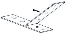
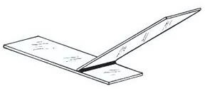
3. Place the edge of the second slide (the spreading slide) into the drop of bacteria and stain until it spreads along the entire edge.
4. Push the spreading slide along the bottom slide, dragging the stain and bacteria evenly.
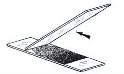
5. Air dry.
6. Examine under the microscope, and show your instructor at 1000x magnification.
7. After your instructor’s approval of your staining technique, dispose of your slides in the plastic beaker labeled “Self prepped” in the Discard Area.
REFERENCES
Brown, A. E. (2009). Benson’s Microbiological Applications: Laboratory Manual in General Microbiology. New York: McGraw Hill.
Chess, B. (2015). Laboratory Applications in Microbiology: A Case Study Approach. New York: McGraw Hill.
Microscope Slide Technique. (2017, December). Retrieved from Microscope Science:http://www.microscopescience.com/microscope-slide-techniques-bacterial-morphology/
Pommerville, J. (2007). Preparation of a Bacterial Smear and the Simple Stain Technique. In J. Pommerville, Alcamo’s Laboratory Fundamentals of Microbiology (p. Chapter 4). Boston: Jones & Bartlett, LLC. Retrieved from http://samples.jbpub.com/9780763795573/95573_ch04.pdf
