8 Endospore Stain
The endospore stain differentiates between developmental stages of bacteria. There are two genera of bacteria, Clostridium and Bacillus, that make up most of the Firmicutes phylum. They have two stages of development: vegetative stage and sporulation stage. The vegetative stage is when the bacterial cell is the normal actively growing vegetative cell. It is capable of metabolism, enzymatic activity, and has the normal water content. Due to actively reproducing, its DNA is exposed, and also has sensitivities to extreme temperatures, radiation, and chemicals. The sporulation stage is when the vegetative cell is forming endospores as insurance for when essential nutrients may be depleted, especially when carbon and nitrogen become unavailable. Endospores are a resting, or dormant, stage that protect DNA and proteins from unfavorable conditions. They are dehydrated structures that do not grow or carry out metabolism, so they are insensitive to antibiotics. They have many layers surrounding the core, which contains the DNA. The outermost layer is made up of a thick protein coat of exosporium that forms a protective barrier. (Figure 8.1). Endospores are resistant to heat, radiation, acids, and many chemicals due to the composition of these layers.
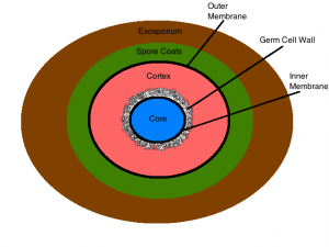
Figure 8.1 This drawing illustrates the structure of an endospore and its layers of protection.
https://microbewiki.kenyon.edu/index.php/File:Figure_3_endospore_structure.png
MATERIALS
Each student should have:
Blue rack
2 glass slides
Malachite green stain
Stain bottle rack: safranin
Lens paper
Windex (depends on instructor)
Inoculating loop
Wax pencil
Metal slide clip
Bunsen Burner
Striker
1 slant culture of Bacillus subtilis
Microscope
PROCEDURE OF SMEAR PREPARATION
MAKE 2 SMEAR PREPS. ONE WILL BE USED AS A BACKUP.
- Draw a circle with the wax pencil on a slide.
- Flip the slide over. Write “Up”.
- Place a small drop of distilled water onto the slide within the wax circle.
- Using aseptic technique, remove a small amount of bacteria from a slant
- Air dry the slide. Do not apply heat to a wet slide. Do not blow on the slide.
- Heat fix the slide.
PROCEDURE OF ENDOSPORE STAIN
STAIN ONLY ONE SLIDE. You may not need the second slide. This serves as a backup in case you have error in your staining procedure. It saves you time.
- Place one smear prep on a boiling hot water bath.
- Place a piece of paper towel over smear.
- Saturate towel & slide with malachite green for 5 minutes.
- Decolorize slide with distilled water until clear over stain tray.
- Counterstain with safranin for 60 seconds.
- Rinse with distilled water.
- Blot dry with a paper towel.
- Examine under the microscope, and show your instructor at 1000x magnification.
- After your instructor’s approval of your staining technique, dispose of your slides in the plastic beaker labeled “Self prepped” in the Discard Area.
REFERENCES
Brown, A. E. (2009). Benson’s Microbiological Applications: Laboratory Manual in General Microbiology. New York: McGraw Hill.
Chess, B. (2015). Laboratory Applications in Microbiology: A Case Study Approach. New York: McGraw Hill. Microbe Wiki. (2012, December 16). Retrieved from Bacterial Endospores:
https://microbewiki.kenyon.edu/index.php/Bacterial_endospores
OpenStax. (2016, November 1). Microbiology. Retrieved from https://legacy.cnx.org/content/col12087/1.4
