
Lab 12: Metabolism and Digestive Physiology
Background
Digestion is the process of breaking down food into smaller molecules that the body can absorb for energy, growth, and repair. This is accomplished through two main types of digestion: mechanical, which involves physical breakdown like chewing, and chemical, which uses enzymes and acids to break down food. The process starts in the mouth and continues through the esophagus, stomach, and intestines, with the help of organs like the liver and pancreas.
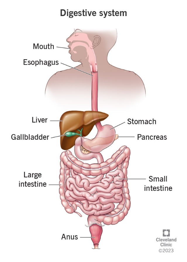
Key organs involved
Mouth: The start of digestion, where food is mechanically broken down by chewing and chemically broken down by saliva.
Esophagus: A tube that carries food from the mouth to the stomach.
Stomach: A muscular organ that mixes food with digestive juices and acids to break it down further.
Small intestine: The primary site for nutrient absorption and further chemical digestion.
Large intestine: Absorbs water from the remaining indigestible food matter.
Liver and pancreas: These organs produce enzymes and other substances (like bile) that are crucial for chemical digestion in the small intestine.
Objectives
- To list the digestive system enzymes involved in the digestion of proteins, fats, and carbohydrates; to state their site of origin; and to summarize the environmental conditions promoting their optimal functioning.
- To recognize the variation between different types of enzyme assays.
- To name the end products of digestion of proteins, fats, and carbohydrates.
- To perform the appropriate chemical tests to determine if digestion of a particular foodstuff has occurred.
- To cite the function(s) of bile in the digestive process.
- To discuss the possible role of temperature and pH in the regulation of enzyme activity.
Salivary Digestion of Carbohydrates
Digestion of Carbohydrates begins in mouth where the salivary glands secrete an enzyme, amylase that begins the hydrolysis of complex polysaccharides:
Plant starches or animal glycogen ——— >Disaccharides (Maltose, sucrose, lactose)
Salivary amylase has an optimum pH of around 6.8, which is roughly pH found in the mouth.
In the following experiment, we will examine amylase digestion of starch, using Benedict’s test to measure maltose formation and Lugol’s (iodine) solution to test for starch.
Experimental Procedure:
- Collect 7 ml of your own saliva from a dedicated member of the group in a graduate cylinder (not including the bubbles). Dilute saliva with an equal amount of distilled water to bring this to a 14 ml total volume solution.
- Test the saliva with pH paper. Is it acid or alkaline?
- Prepare and label four test tubes as follows (Use 0.5% starch solution):
Tube 1: 3 ml. of starch + 3ml. of water. Incubate at 37 C. Tube 2: 3 ml. of starch + 3ml. of saliva. Incubate at 37 C. Tube 3: 3 ml. of starch + 3ml. of saliva. Incubate on ice. Tube 4: 3 ml. of starch + 3ml. of saliva + 5 drops of concentrated HCl. Incubate at 37 C.
Incubate all test tubes for 1 hour.
4. Starch Test: Take 5 drops of incubate on a white tile and add 1 drop of Lugol’s iodine. A dark purple color indicates presence of starch. Shades of reddish brown indicate lesser amount of starch. (+++), (++), (+) (-)
5. Maltose Test: Add 4 ml. of Benedict’s solution and place in a boiling water bath for 2 mins. Compare the color developed. (+++) red; (++) orange; (+) green; (-) blue {PLACE IN THE SAME ORIGINAL TEST TUBE}
Gastric digestion of Proteins
Protein digestion begins in the stomach where the enzyme splits proteins into shorter polypeptide chains containing amino acids. Secretion and activation of pepsin occurs as follows: Chief cells in gastric pit —–> Pepsinogen + HCl —— >Pepsin
Experimental Procedure:
-
- Place 3 ml. of albumin soln. (5%) in 4 test tubes.
- Add the following solutions to the tubes:
Tube 1: 5 ml pepsin (5% solution) + 5 ml. HCl Tube 2: 5 ml pepsin (5% solution) + 5 ml. water Tube 3: 5 ml water + 5 ml. HCl Tube 4: 5 ml pepsin (5.0 % solution) + 5 ml. NaOH (0.5 %)
3. Allow the tubes to incubate at 37 C for 1 hour. Test the final pH of the solution and estimate the amount of proteins digested using a scale of (+++), (++), (+), (-) to compare the four tubes.
Digestion of fat with pancreatic lipase and bile salts
Pancreatic lipase has a major role in fat digestion, but by itself lipase is ineffective, because it is a water-soluble enzyme trying to act on large lipid droplets, which are insoluble. Bile salts help overcome this problem by emulsifying fat into smaller droplets, so that lipase has a larger surface area for the hydrolysis of fats.
Pancreas also aids digestion by secreting sodium bicarbonate. This compound provides a pH around 7.8 in the small intestine, which is optimal for the action of pancreatin enzymes. In the following exercise, we will examine some aspects of the action of pancreatic lipase and bile salts on lipids.
Effects of Bile salt on Oil:
-
- In each of two test tubes (A and B) place 3 ml of distilled water and 3 ml of vegetable oil.
- To tube B add a small pinch of bile salts. Shake each tube for 30 secs. And observe it for several mins.
Effects of pancreatic enzymes:
3. Add litmus powder to dairy cream gradually until a light purple color is produced. Pre-incubate the litmus cream and a 1% pancreatin solution (tubes 1 and 3 from the table below) at 37 C for 5 mins.
This ensures the enzyme and substrate (fat in cream) are at body temperature before mixing. The basis of this assay is a pH change that is detected by a litmus powder indicator. Alkaline or neutral solutions containing litmus are light purple but will turn reddish pink in the presence of acid. Since fats are digested to fatty acids (organic acids) during hydrolysis, they lower the pH of the sample they are in. Litmus cream (fresh cream providing the fat substrate to which litmus powder was added) will turn from a purple color to pink if the solution is acidic.
4. Prepare a series of tubes as follows:
| Tube 1: | 3 ml cream + 3 ml pancreatin |
| Tube 2: | 3 ml cream + 3 ml water |
| Tube 3: | 3 ml cream + 3 ml pancreatin + pinch of bile salts |
| Tube 4: | 3 ml cream + 3 ml water + pinch of bile salts |
5. Incubate all tubes in 37 C water bath for 1 hour or until a color change occurs in one tube. Blue litmus will turn pink in an acid environment. Test the pH using pH paper and note the odor of each tube.
DIGESTION
Carbohydrates:
Mouth:
Salivary Amylase
Polysaccharides ————————> Disaccarides, Trisaccharides and dextrins
Duodenum (Pancreatic juice):
Pancreatic Amyalse
Polysaccharides ————————————-> Disaccarides and Trisaccharides
Intestine (Brush border Enzymes):
Dextrinase
dextrins ————————————–> single dextrose (glucose)
Maltase
Maltose —————————————– >2 molecules of glucose
Sucrase
Sucrose —————————————– >glucose + fructose
Lactase
Lactose ——————————————> glucose and galactose
Proteins:
Stomach:
Pepsin*
Proteins ——————————————> Peptides
Duodenum (Pancreatic juice):
Pancreatic Trypsin^
Proteins ——————————————> Peptides
Chymotripsin@
Proteins ——————————————> Peptides
Carboxypeptidase
Peptides —————————————– > removes –COOH group from peptide
Intestine:
Aminopeptidase
Peptides —————————————– > removes amino (NH2) group
Dipeptidase
Dipeptides ————————————–> Single amino acids
HCl
*Pepsinogen (Inactive) ————————————————– >Pepsin*
Enterokinase (from intestine)
^ Trypsinogen (Inactive) ————————————————- >Trypsin^
Trypsin
@Chymotrpsinogen (Inactive) ——————————————> Chymotrypsin@
Lipids:
Mouth:
Lingual Lipase
Triglycerides —————————————–> Fatty acids and monoglycerides
Intestine: (Bile and Pancreatic juice)
Bile Salts
Large Triglyceride mols. —————————— >Broken into smaller mols.
Pancreatic Lipase
Triglycerides ——————————————> Fatty acids and monoglycerides
Hormones of GI tract
| Hormone | Origin | Effects |
| 1. Gastrin | Pyloric Stomach | 1. Stimulates gastric secretion and its motility |
|
2. Gastric Inhibitory Peptide (GIP) |
Duodenal mucosa | 1. Inhibits gastric secretion and its motility
2. Stimulates release of insulin |
| 3. Secretin | Duodenal mucosa | 1. Inhibits gastric secretion and its motility
2. Stimulates release of HCO3 from pancreas 3. Increases rate of bile secretion |
| 4. Cholecystokinin | Duodenal mucosa | 1. Inhibits gastric secretion and its motility
2. Causes contraction of gall bladder 3. Stimulates secretion of pancreatic enzymes |