
Lab 8: Crayfish Heart & Blood Physiology
Materials needed for blood physiology activity:
Fan for blood drying
Glass slides
Giemsa staining solution (mix 1 part Giemsa stock solution with 9 parts phosphate buffer)
Compound light microscopes
Methanol
pH 7.2 phosphate buffer
Immersion oil
5 µl microcapillary with bulb for collecting crayfish hemolymph (one per crayfish)
Coplin staining jar (2 units)
Background
Cardiac muscle contraction is an involuntary process driven by electrical signals from the sinoatrial (SA) node that causes coordinated heart muscle cell (cardiomyocyte) contractions. This process, called excitation-contraction coupling, begins when an electrical action potential opens L-type calcium channels, allowing calcium to enter the cell. This initial calcium influx then triggers a larger release of calcium from the sarcoplasmic reticulum through the calcium-induced calcium release (CICR) mechanism. The increased intracellular calcium binds to troponin, causing the protein to move and expose binding sites on actin filaments, allowing myosin heads to bind and slide the filaments to shorten the sarcomere and create muscle contraction.
The process of cardiac muscle contraction:
- Electrical impulse: An electrical action potential is generated by the SA node and spreads through the heart via gap junctions in specialized conductive fibers.
- Calcium influx: The action potential travels along the cardiomyocyte’s cell membrane (sarcolemma) and down the T-tubules, triggering L-type calcium channels to open and allowing calcium from the extracellular fluid to enter the cell.
- Calcium-induced calcium release (CICR): The small amount of incoming calcium activates ryanodine receptors on the sarcoplasmic reticulum, causing a much larger release of stored calcium into the cytoplasm.
- Crossbridge cycling: The surge of calcium binds to troponin C, causing it to move tropomyosin and expose binding sites on actin filaments. Myosin heads then bind to these sites, and using ATP, they pull the actin filaments toward the center of the sarcomere, resulting in muscle contraction.
- Relaxation: The action potential ends, calcium influx stops, and the cell actively pumps calcium back into the sarcoplasmic reticulum and out of the cell. This reduces the intracellular calcium concentration, causing calcium to detach from troponin, which allows tropomyosin to cover the actin binding sites again and the muscle to relax.
In this lab, you will measure the crayfish heartbeat and understand how it can be modulated by drugs acting on the nervous system. In addition, you will conduct a blood smear to inspect the cell contents of the hemolymph and compare these with the cell contents of blood.
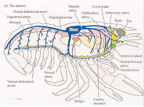
Crayfish blood analysis
-
- Collect a blood sample from the caudal blood vessels or by cutting the tail when you are preparing the crayfish for the heart activity.
- Place a drop of blood on a clean microscope slide.
- Spread the drop into a thin smear using another slide held at a 45-degree angle.
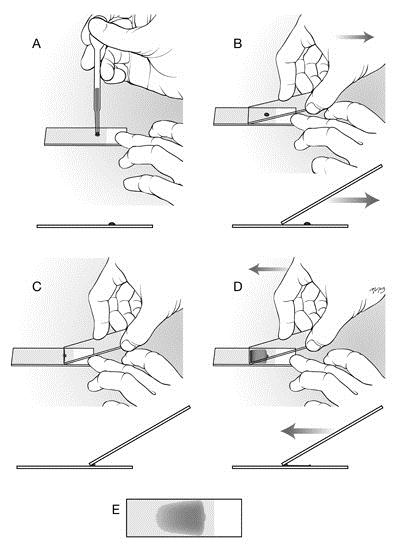
4. Blood smearing technique using two glass slides.
5. Set this aside to allow the smear to air dry for 30 min.
6. Fix the slides by immersing them in methanol for 1 minute. Let it air dry for 30 seconds.
7. Place the slides in the working Giemsa solution for 20-30 minutes. Place the slide in a staining jar and flood the smear with the prepared Giemsa stain solution.
8. Gently rinse the slide by dipping it in pH 7.2 phosphate buffer, followed by a rinse with distilled or neutral water. Be careful not to wash away the smear.
9. Stand the slide upright and let it air dry completely for 10 min. Once dry, add a drop of immersion oil and examine the stained hemolymph cells under a microscope with the 100x oil immersion lens.
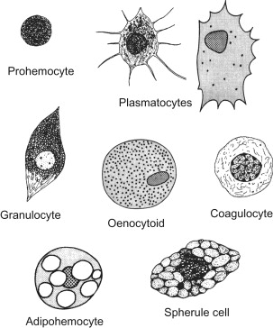
|
Prohemocytes |
– Small, rounded cells – Large nucleus and basophilic cytoplasm – Considered immature or precursor cells that can differentiate into other hemocyte types |
|
Plasmatocytes |
– Most common type of hemocyte – Spindle or leaf-like shape with long, hair-like projections (pseudopodia or filopodia) – Actively involved in phagocytosis, encapsulation, and nodulation |
|
Granular Hemocytes (Granulocytes) |
– Round or oval with numerous cytoplasmic granules – Possess macrophage-like functions, including phagocytosis and forming “sticky nets” to trap pathogens – Involved in cellular immune responses like encapsulation and nodulation |
|
Coagulocytes |
– Contain cytoplasmic granules – Their main function is to participate in the clotting of hemolymph to seal wounds |
|
Oenocytoids |
– Large, often irregular or round in shape – Typically have a round, “fried egg” appearance with a large nucleus and cytoplasmic granules – Not involved in phagocytosis – Function is not fully understood, but they are involved in some immune responses |
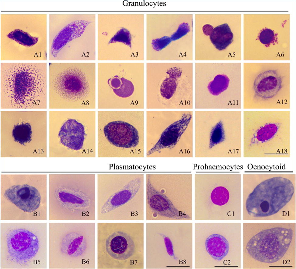
Use the pictures and descriptions above as guides. Draw and label what you see underneath the microscope in your lab report, while also including the total magnification you are viewing the blood sample at.
The Crayfish Heart Activity
Background
Vertebrate hearts are myogenic; each muscle cell has intrinsic pacemaker properties. Each cell will beat in the absence of any neural input. The cells throughout the heart beat in a coordinated fashion because they are electrically coupled and beat at the same rate as the pacemaker cells in the sino-atrial and atrial-ventricular nodes. In most vertebrates, the blood is pumped into a closed circulatory system of arteries and veins.
Many invertebrates, including crustaceans like the crayfish, have neurogenic hearts. The myocardial cells will not beat without neural input from the cardiac ganglion. The resting heart rate and contractile force are set by this neural input. Crayfish also have an open circulatory system. Blood flows into the heart through dorsal ostia, and is pumped into body sinuses through arteries at both the posterior and anterior ends of the ventricle. The beats of both myogenic and neurogenic hearts are modulated by neurotransmitters. Vertebrate hearts are excited by epinephrine and inhibited by acetylcholine. In living crayfish, cardioexcitatory peptides increase the heart’s rate and contractile force, while GABA inhibits both rate and force. Biogenic amines like Serotonin and Dopamine also have effects on the crayfish heartbeat. In this laboratory exercise, students will use a force transducer to monitor the contractility of the crayfish heart as it is subjected to various imposed conditions, such as: the effect of adding Serotonin and GABA to change the heart rate and contractile force of the exposed heart; and the effect of cold temperature on cardiac muscle activity.
The Dissection
1. Cover a crayfish with ice for 5 minutes.
2. With scissors, remove the claws, walking legs, and abdomen (“tail”) from the crayfish. Collect the hemolymph for the blood smear.
3. The crayfish heart is located dorsally, at the posterior end of the thorax. Use the scissors to carefully remove a small section of carapace over the heart. The hypodermis, the red membrane directly beneath the carapace, should also be removed.
4. The beating yellow-white ventricle of the heart should be clearly visible.
5. Use four #2 insect pins to pin the crayfish securely into a wax or Sylgard dish.
6. Cover the crayfish with 100 ml of room temperature Ringer’s. If 100 ml of saline doesn’t cover the crayfish, use more. It is important to keep track of exactly how much saline is in the dish.
The Preparation
1. Place the dissection dish so that the crayfish ventricle is directly below the end of the
transducer. The force transducer should be about 15 cm above the heart, with the blade of the transducer being horizontal (Figure AM-7-L1).
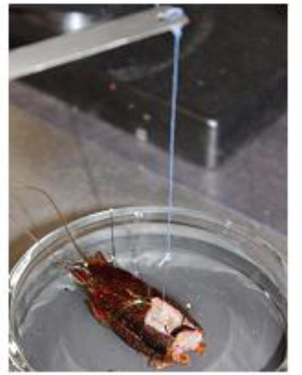
Figure AM-7-L1: The crayfish heart preparation
2. Bend a metal pin to form a hook. Tie a 20 cm length of thread behind the head of the hook.
3. Push the hook through the ventricle wall until the bend of the hook is inside the heart.
4. Tie the loose end of the thread to the hole in the blade or on the hook of the transducer. Loosen the clamp holding the transducer and gently raise it on the ring stand. Put enough tension on the thread to raise the ventricle very slightly. (Figure AM-7-L2).
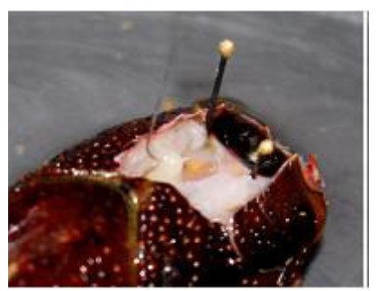
Figure AM-7-L2: Force transducer hook placed through the ventricle of the heart.
Warning: The heart preparation used in this experiment is functional for a limited period of time. Keep the heart muscle covered in saline. To conserve time, complete all the exercises in the experiment before analyzing the data.
Exercise 1: The Heart Rate
Aim: To record the mechanical trace produced by the contraction of a resting heart, and to determine the resting heart rate.
Approximate Time: 30 minutes (including dissection)
Procedure
1. Type Resting in the Mark box to the right of the Mark button.
2. Click the Record button and press the Enter key on the keyboard to attach the comment to the record. Click AutoScale to increase the size of the deflection on the Main window.
3. Record the heart contractions for thirty seconds. A sample recording can be seen in Figure AM- 7-S3.
4. Click Stop to halt the recording.
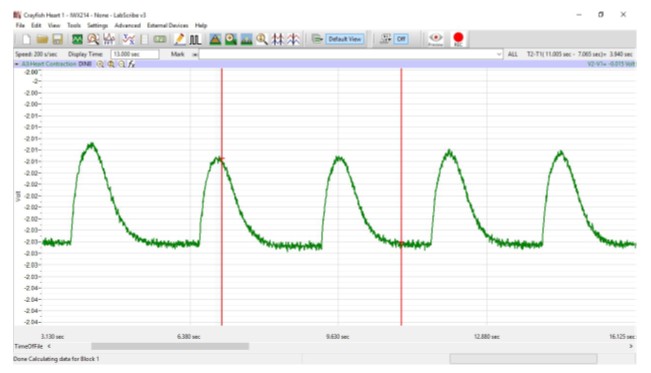
Figure AM-7-S3: Recording of the contractions of the crayfish heart.
5. Select Save As in the File menu, type a name for the file. Choose a destination on the computer in which to save the file, like your lab group folder. Designate the file type as *.iwxdata. Click on the Save button to save the data file.
Exercise 2: Effects of Cold Temperature
Aim: To record changes in heart rate after the heart is bathed in cold Ringer’s solution.
Approximate Time: 15 minutes
Procedure
1. Type Room Temp Ringer’s in the Mark box to the right of the Mark button.
2. Click the Record button and press the Enter key to attach the comment to the recording. Click AutoScale to increase the size of the deflection on the Main window.
3. Record the heart contractions for thirty seconds.
4. Click Stop to halt the recording.
5. Draw off the room temperature Ringer’s with a syringe, and add chilled saline, taking care to replace the saline with the same volume as was drawn off.
Warning: It is important to keep the saline at the same depth throughout the experiment, as the depth of the saline will affect the amplitude of the recorded beats. The dissecting dish should also be firmly affixed to the table with clay so that it won’t be accidentally bumped, changing the tension on the ventricle.
6. Type Cold Ringer’s in the Mark box.
7. Click the Record button and press the Enter key to attach the comment to the recording.
8. Record until the heart has recovered from the effects of cold Ringer’s solution.
Note: Recovery is when the amplitude and rate of the heart contraction have returned to the resting values.
9. Click Stop to halt the recording.
10. Select Save in the File menu.
Exercise 3: Effects of Drugs
Aim: To monitor the effects of Serotonin and GABA on the amplitude and rate of heart contraction. Approximate Time: 45 minutes
Procedure-Serotonin
1. Type Pre-Serotonin control in the Mark box to the right of the Mark button.
2. Click the Record button. Press the Enter key on the keyboard to mark the recording. Click AutoScale to increase the size of the deflection on the Main window.
3. Record the heart contractions for thirty seconds.
4. Type Serotonin 10-6M in the Mark box to the right of the Mark button.
5. Add 100 microliters of the 10-3M stock Serotonin solution to the saline in the dish. Gently stir the saline to disperse the Serotonin.
Note: If you used a volume other than 100 ml, adjust the amounts of the stock solution accordingly. For example, if you covered the crayfish with 150 ml, add 150 microliters of the stock Serotonin solution to create a 10-6M Serotonin solution.
6. Click Record to start the recording, and press the Enter key on the keyboard.
7. Record the effects of 10-6M Serotonin for two minutes.
8. Click Stop to halt the recording.
9. Add an additional 900 microliters of the 10-3M Serotonin solution to the saline in the dish to create a 10-5M solution. Gently stir the saline to disperse the Serotonin throughout the saline.
10. Type Serotonin 10-5M in the Mark box to the right of the Mark button.
11. Click Record to start recording and press the Enter key.
12. Record for two minutes.
13. Click Stop to halt the recording.
14. Add 10 ml of the 10-3M Serotonin stock solution to the saline in the dish to create a 10-4M Serotonin solution.
15. Type Serotonin 10-4M in the Mark box to the right of the Mark button.
16. Click Record to start recording and press the Enter key to attach the comment to the recording.
17. Record for two minutes.
18. Click Stop to halt the recording.
19. Replace the saline with 100 ml of fresh saline, and allow the heart to recover.
Note: It is possible that the heart will not return to pre-treatment conditions. In this case, wait until the heart has come to a new steady-state, and record the heartbeat at that time. This should occur within ten minutes after the saline change.
20. Type Post-Serotonin recovery in the Mark box.
21. Once the heart has recovered, click Record to start the recording and press the Enter key.
22. Record for thirty seconds.
23. Click Stop to halt the recording.
24. Select Save in the File menu.
Procedure-GABA
1. Type Pre-GABA control in the Mark box to the right of the Mark button.
2. Click Record to start the recording and press the Enter key to attach the comment to the recording.
3. Record for thirty seconds.
4. Click Stop to halt the recording.
5. Add 100 microliters of the 10-3M stock GABA solution to the saline in the dish. Gently stir the saline to disperse the GABA throughout the saline.
6. Type GABA 10-6M in the Mark box to the right of the Mark button.
7. Click the Record button and press the Enter key.
8. Record the heart contractions for two minutes.
9. Click Stop to halt the recording.
10. Add an additional 900 microliters of the 10-3M GABA solution to the saline in the dish to create a 10-5M solution. Gently stir the saline to disperse the GABA throughout the saline.
11. Type GABA 10-5M in the Mark box to the right of the Mark button.
12. Click Record to start recording and press the Enter key.
13. Record for two minutes.
14. Click Stop to halt the recording.
15. Add 10 ml of the 10-3M GABA stock solution to the saline in the dish to create a 10-4M GABA solution. Gently stir the saline to disperse the GABA.
16. Type GABA 10-4M in the Mark box to the right of the Mark button.
17. Click Record to start recording and press the Enter key to attach the comment to the recording.
18. Record for two minutes.
19. Click Stop to halt the recording.
20. Replace the saline with 100 ml of fresh saline, and allow the heart to recover.
21. Type Post-GABA recovery in the Mark box.
22. Once the heart has recovered, click Record to start the recording and press the Enter key.
23. Record for thirty seconds.
24. Click Stop to halt the recording.
25. Select Save in the File menu.
Record your data in the data tables for each exercise in the lab report. Make sure to answer the questions at the end of the lab report as well.