
Lab 5: Introduction to Organismal Systems
Disclaimer: This lab will involve the dissection of once live organisms, and at times, this practice has been controversial, but these organisms have been humanely euthanized and preserved with educational purposes in mind. In order to facilitate the accurate learning of anatomical structures, it is necessary to observe and dissect an actual specimen as opposed to performing a virtual dissection. If, at any time, you are not feeling well, you can always step out of the lab and take a break. If you are adverse to doing the cutting, then pair up with someone who doesn’t mind, you will however, still be responsible for observing and participating in the lab activity.
Purpose: In this lab, you will dissect an earthworm, yellow perch bony fish, and a rat in order to observe their external and internal structures. After identifying the external and internal structures, compare them across the organisms and consider how they have been adapted of a “design” that is particularly suited for the habitat that they live in.
Think about the following questions as you conduct your dissections today. How are the organismal structures similar? How are they different? How are the structures linked to their function? How have they been altered through evolutionary history? Which structures are similar to humans? How are they different?
Directions: Once you have identified all of the external structures for each organism have your instructor quiz you on them, then move onto the internal structures and repeat the same process before moving onto the next organismal dissection. Make drawings of the external and internal anatomy, with labels, of each organism, to be graded as your lab report.
Dissection of Earthworm (annelid)
Background: Among the most familiar invertebrate animals are the earthworms, members of the phylum Annelida. The word annelida means “ringed” and refers to a series of rings or segments that make up the bodies of the members of this phylum. Internally, septa, or dividing walls, are located between the segments. There may be more than 100 segments in an adult worm. http://en.wikipedia.org/wiki/Earthworm
Digestive System: The earthworm takes in a mixture of soil and organic matter through its mouth, which is the beginning of the digestive tract. The mixture enters the pharynx, which is located in segments 1–6. The esophagus, in segments 6–13, acts as a passageway between the pharynx and the crop. The crop stores food temporarily. The mixture that the earthworm ingests is ground up in the gizzard. In the intestine, which extends over two-thirds of the body length, digestion and absorption take place. Soil particles and undigested organic matter pass out of the worm through the rectum and anus.
Reproductive system: The clitellum is a swelling of the body found in sexually mature worms and is active in the formation of an egg capsule, or cocoon. Eggs are produced in the ovaries and pass out of the body through female genital pores. Sperm are produced in the testes and pass out through tiny male genital pores. During mating, sperm from one worm travel along the sperm grooves to the seminal receptacles of another worm. Fertilization of the eggs takes place outside the body as the cocoon moves forward over the body, picking up the eggs of one worm and the sperm of its mate.
Circulatory system: The pumping organs of the circulatory system are five aortic arches. Circulatory fluids travel from the arches through the ventral blood vessel to capillary beds in the body. The fluids then collect in the dorsal blood vessel and reenter the aortic arches.
Nervous system: It consists of the ventral nerve cord, which travels the length of the worm on the ventral side, and a series of ganglia, which are masses of tissue containing many nerve cells. The nerve collar surrounds the pharynx and consists of ganglia above and below the pharynx. Nervous impulses are responsible for movement and responses to stimuli. Each segment contains an enlargement, or ganglion, along the ventral nerve cord. Excretory functions are carried on by nephridia, which are found in pairs in each body segment. They appear as tiny white fibers on the dorsal body wall. The earthworm has no gills or lungs. Gases are exchanged between the circulatory system and the environment through the moist skin.
Procedure:
1. Place an earthworm in the dissecting tray & rinse off the excess preservative. Identify the dorsal side, which is the worm’s rounded top, and the ventral side, which is its flattened bottom. Turn the worm ventral side up, as shown in the diagram below.
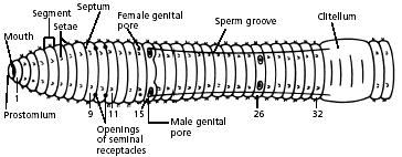
2. Use a hand lens as you observe all parts of the worm, externally and internally. Find the anterior end by locating the prostomium, which is a fleshy lobe that extends over the mouth. The other end of the worm’s body is the posterior end, where the anus is located.
3. Locate the clitellum, which extends from segment 33 to segment 37. Look for the worm’s setae, which are the minute bristle-like spines located on every segment except the first and last one.
4. Refer again to the diagram of the ventral view of the worm to locate and identify the external parts of its reproductive system. Find the pair of sperm grooves that extend from the clitellum to about segment 15, where one pair of male genital pores is located. Look also for one pair of female genital pores on segment 14. There is another pair of male genital pores on about segment 26. Try to find the two pairs of openings of the seminal receptacles on segment 10. Note: These openings are not easy to see.
5. Turn the worm dorsal side up. Using a scalpel and scissors, make a shallow incision in the dorsal side of the clitellum at segment 33. CAUTION: Scalpels and scissors are very sharp. Report any cuts to your teacher. Using the forceps and scalpel, spread the incision open, little by little. Separate each septum from the central tube using a dissecting needle, and pin down each loosened bit of skin. Continue the incision forward to segment 1.
6. Use the diagram below to locate and identify the five pairs of aortic arches, or hearts. Then find the dorsal blood vessel. Look for smaller blood vessels that branch from the dorsal blood vessel.
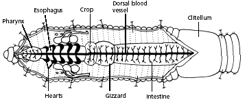
7. Locate the digestive tract, which lies below the dorsal blood vessel. Refer to the diagram above to locate the pharynx, esophagus, crop, gizzard, and intestine.
8. To find organs of the nervous system, push aside the digestive and circulatory system organs. Use the diagram below to locate the ventral nerve cord. Trace the nerve cord forward to the nerve collar, which circles the pharynx. Find one pair of ganglia under the pharynx and another pair of ganglia above the pharynx. The ganglia above the pharynx serve as the brain of the earthworm.
9. The worm’s excretory organs are tiny nephridia. There are two in every segment. Use the preceding diagram to locate some nephridia.
10. Clean up your work area and wash your hands before leaving the lab.
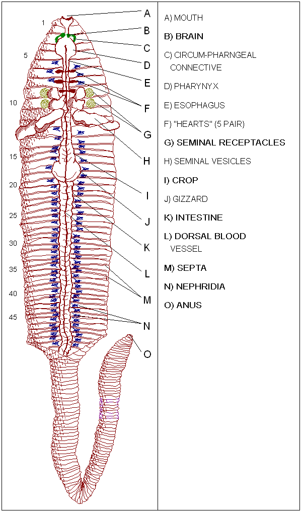
From: http://sps.k12.ar.us/massengale/earthworm_dissection.htm
Dissection of Yellow Perch (bony fish)
Introduction:
In this lab each student will work with a partner in order to learn from the dissection of a Perch. Dissection gives the student the opportunity to observe the placement of organs and their relationships to one another. Pictures and diagrams will aid in the completion of this lab dissection. Be thorough and do not rush through the lab. Read all directions carefully and make all drawings as accurate and neat as possible.
Materials:
Preserved perch
Dissecting tray
Dissecting probe and scissors
Procedure
A. External Anatomy of Perch:
-
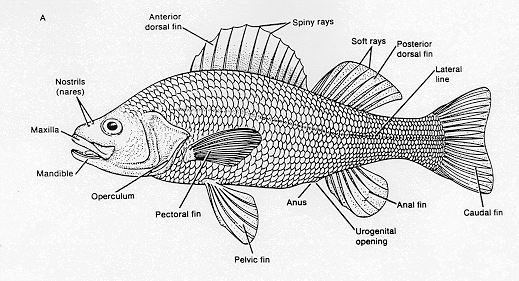
- Place a preserved perch on a dissecting tray. Locate the head region. Examine the eyes.
- Are there any eyelids present? _______.
- Examine the 5 types of fins. Each fin has a purpose or job, what is the purpose of the following fins?
a). caudal fin_________________
b). dorsal fins________________
c). pectoral fin _______________
d). anal _________________
e). pelvic ________________
4. Locate the lateral line. Using the hand lens and look at the line and the surrounding area.
B. Internal Anatomy of Perch:
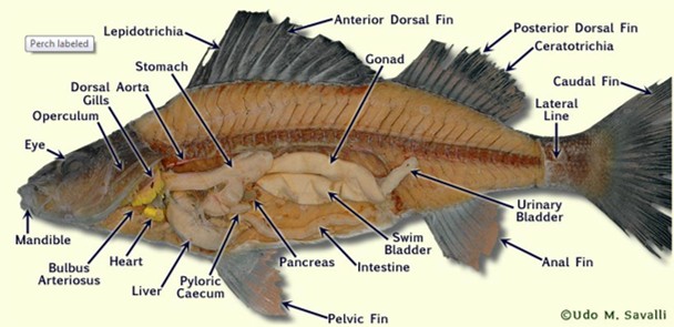
5. Using your thumb, lift up the edge of the operculum and raise it up as far as you can. Using your scissors, cut the operculum off as close to the eye as possible. You have exposed the gills. The gill are layered one on top of another. Using your probe, carefully lift each of these layers.
6. How many layers do you find? ___________
7. Using your scissors, remove one of these layers. Examine the feathery structure.
8. To expose the internal organs you will cut away part of its muscular wall. Grasp your fish, holding it with your thumb on one side and fingers on the other. Turn your hand upward to expose the ventral surface. Using your scissors, insert the point into the skin just in front of the anus. Cut forward to the gills. Be careful not to destroy any of the internal organs since they are mostly found in this area.
9. The fish contains a 2 chambered heart. Locate this organ found just behind and below the gills.
Label the parts of the heart below.
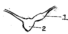 Fish Heart
10. Part 1 _______________ Function: _________________
11. Part 2 _______________ Function: _________________
12. Locate the tube-like digestive system. Begin just behind the mouth in the area called the pharynx. This area leads into the gullet or the opening of the esophagus. The esophagus leads into the stomach. Cut out the stomach and split it open.
13. Locate the rather large liver located just in front of the stomach.
14. Try to locate the kidneys, found just below the spinal column. Their main function is to rid the body of nitrogenous waste.
15. The swim bladder is the last remaining organ to be identified. It is located between the kidneys and gonads.
16. What is the function of the swim bladder? _____________________________________
Dissection of Rat (mammal)
Introduction:
Each student will work with a partner to learn about the anatomy and physiology of a rat. Dissection of the rat offers the opportunity to compare the anatomical differences between the annelid and bony fish to the mammal. The rat serves as a model of anatomical and physiological similarities to humans. Be thorough in following directions of this dissection.
Materials:
Preserved rat
Dissecting tray
Dissecting probe and scissors
Procedure
A. External Anatomy of Rat:
1. Place the preserved rat specimen in a dissecting tray, ventral side up. Locate the following regions of the body: cranial, cervical, pectoral, thorax, abdomen, pelvic and caudal regions. Identify external features.
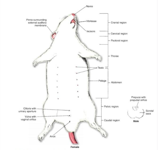 2. Determine the sex of the rat specimen by examining the pelvic region. State whether the specimen is male or female.
B. Internal Anatomy of Rat
4. Tie one end of a piece of string around the right front paw, wrap the string around the outside of the dissecting, and tie the other of the string to the left front paw. Do the same for the hind paws.
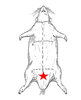
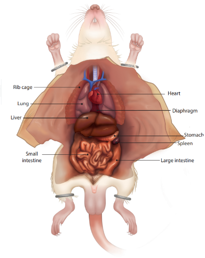
5. Using the below image as a guide, make cuts starting at the star and move upward toward the cranial region. Open up the pectoral, thorax, and abdominal regions by pinning the flaps to the dissecting tray.
6. Using the below image, examine and identify the digestive organs. Notice the scattered cream colored fat deposits. Note the size of the liver in proportion to the abdomen.
7. Remove the digestive system by cutting the esophagus above its connection with the stomach (below the diaphragm) and by cutting the rectum. Notice the way the small intestine and large intestine are twisted over one another.
8. Remove the liver carefully and compare the color of the spleen and liver. Note there is no gall bladder in the rat.
9. Observe the exposed kidneys and associated urinary organs, and the reproductive system left behind in the abdominal cavity.
10. Examine the thoracic (pectoral) region by cutting away the ribs to reveal the heart surrounded by a thin membrane called the pericardium, the whitish thymus gland that lies directly over the upper part of the heart and the lungs that lay either side of the heart.
11. Remove the heart and thymus to locate the trachea, which is identifiable by its rings of cartilage. The esophagus lies just underneath the trachea.
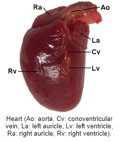
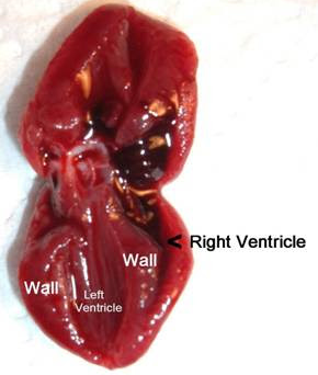
12. Make a coronal incision into the heart (divides the heart into anterior and posterior regions) to view the 2 ventricles. Note the heart of the rat has 4 chambers: 2 atria and 2 ventricles.
Dispose of your rat and its removed parts by placing in the bagged bucket. Clean up your work area by washing dissecting tools and tray.