9 Human Reproduction and Development
Learning Objectives
- Identify and describe the functions of the major structures of the male reproductive system.
- Trace the path of sperm within the male reproductive tract.
- Identify and describe the functions of the major structures of the female reproductive system.
- State the major events during the ovarian cycle.
- Trace the path of an egg from ovary to implantation to birth.
- Explain the process of fertilization.
- Describe the three stages of embryonic development.
- Explain the main events that occur during fetal development.
INTRODUCTION
In the study of biology, the term sex refers to the classification according to the male and female reproductive organs and functions that are derived from the chromosomes. The sex of an individual that has the chromosomes XX is termed female whereas an individual with the chromosomes XY is termed male.
Humans produce offspring by the process of sexual reproduction where the haploid gametes of each sex (sperm and egg) join to form one diploid cell, the zygote, through the process of fertilization. The reproductive system of both males and females consists of the gonads, organs which produce the gametes; ducts to transport the gametes; and specialized structures to facilitate fertilization and, in females, nourish and develop the embryo and fetus.
MALE REPRODUCTIVE SYSTEM
The primary functions of the male reproductive system are to (1) produce and transport the male gametes, the sperm, (2) produce and secrete male sex hormones like testosterone, (3) produce fluids that nourish and support sperm, and (4) deliver sperm into the female reproductive tract for fertilization.
The organs of the male reproductive system include the testes, a duct system (epididymis, ductus deferens, ejaculatory ducts, and urethra), accessory glands (seminal vesicles, prostate gland, and bulbourethral glands) and supporting structures (scrotum and penis). (Figure 9.1)
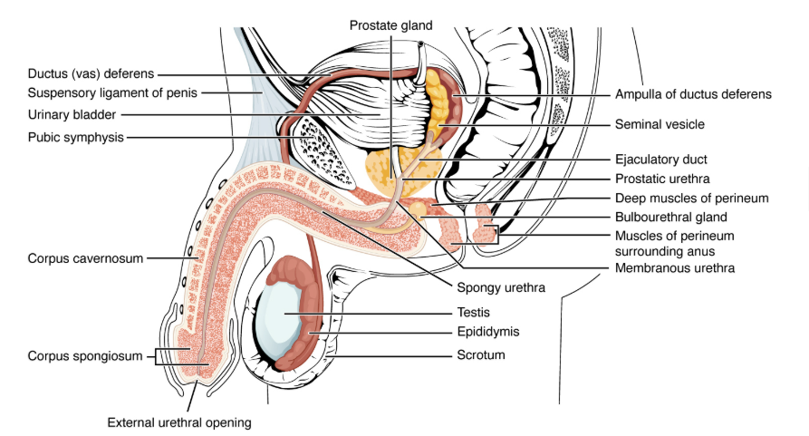
Figure 9.1 The male reproductive system
GONADS
The male gonads are the testes, where sperm and the male sex hormone testosterone are produced. The testes are housed external to the main body cavity in the skin-covered sac of tissue called the scrotum. The scrotum regulates the temperature of the testes as they need to be at a slightly cooler temperature than body temperature to produce sperm. Each testis is divided by septa into lobules. Within the lobules, tightly coiled structures called the seminiferous tubules produce sperm through the process of spermatogenesis (Figure 9.2). These tubules contain spermatogonia that divide to produce primary spermatocytes (sperm), and Sertoli cells that support and nourish the developing sperm.
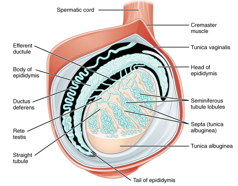
Figure 9.2: Anatomy of the testis
The testes also contain interstitial cells (previously known as Leydig cells), which produce testosterone. Testosterone influences sperm production, development of primary and secondary male sex characteristics, and sexual function. It also regulates bone mass, muscle strength, and fat distribution.
DUCT SYSTEM
After production within the seminiferous tubules of the testes, spermatids (immature sperm) travel to the epididymis, a coiled tube attached to the testis, where they are stored to fully mature to be able to fertilize an oocyte (egg). Mature sperm have a distinctive head, mid-piece and tail region (Figure 9.3). The head of the sperm contains DNA. It is surrounded by the acrosome, which contains enzymes that help it digest and penetrate the protective layers surrounding an oocyte. The mid-piece consists of tightly packed mitochondria, which produce energy for the tail of the sperm, enabling it to swim through the female reproductive tract to reach and fertilize the egg.
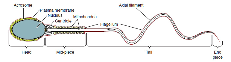
Figure 9.3: Structure of a mature sperm cell
Upon ejaculation, the mature sperm are propelled from the epididymis through the ductus (vas) deferens, long muscular tubes, which eventually join with the seminal vesicles to form the ejaculatory duct, and then the urethra. The male urethra is shared by both the male reproductive system and urinary system. When sperm are released through ejaculation, urine is blocked exiting from the urethra. A muscular bladder sphincter and the nervous system control when urine exits through the urethra.
ACCESSORY GLANDS
As the sperm are propelled through the ductus deferens into the ejaculatory duct, the seminal vesicles contribute fluid that contains large amounts of fructose (energy source for sperm mitochondria). The ejaculatory duct travels through the prostate gland where prostatic fluid is added to the semen. Prostatic fluid contributes to the milky appearance of semen. The prostate gland also produces the enzyme prostate-specific antigen (PSA) that aids in liquefying semen after ejaculation, which is a crucial process for sperm motility. The final addition to seminal fluid is made by the bulbourethral glands that release a thick, salty fluid that lubricates the end of the urethra and the vagina.
PENIS
The penis is the male organ of copulation (sexual intercourse). The shaft of the penis is composed of three cylindrical masses of tissue (two corpora cavernosa and one corpus spongiosum) that become engorged with blood during erection. The penis contains the urethra, which conducts urine or seminal fluid out of the body. It is the copulatory organ of the male, used for sexual intercourse.
Note to students: Write all data and answers to questions on the Lab Report provided.
Activity 1: Identify the Function of the Organs of the Male Reproductive System
Match the male reproductive organ with its function. Record answers on the Lab Report.
|
Term |
Definition |
|
a. Penis |
1. Contributes milky seminal fluid to semen |
|
b. Testis |
2. Expel urine or semen from the body |
|
c. Ductus deferens |
3. Produce sperm |
|
d. Seminal Vesicle |
4. Transport semen from ductus deferens to urethra during ejaculation |
|
e. Ejaculatory duct |
5. Stores sperm for maturation |
|
f. Urethra |
6. Produce thick, salty fluid that lubricates urethra |
|
g. Prostate gland |
7. Muscular tube that transports sperm from epididymis to urethra |
|
h. Epididymis |
8. Copulatory organ |
|
i. Bulbourethral gland |
9. Male gonad |
|
j. Seminiferous tubules |
10. Contributes fructose to semen |
Activity 2: Trace the Path of Sperm through the Male Reproductive System
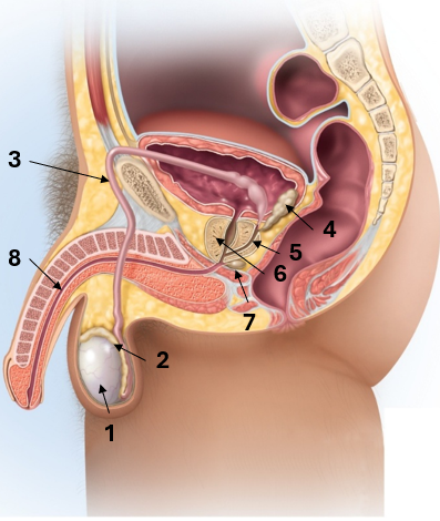
Label male organs in the pathway of sperm from production to ejaculation. Record answers on the Lab Report.
FEMALE REPRODUCTIVE SYSTEM
The primary functions of the female reproductive system are (1) produce ova (eggs) and transport them to the uterus, (2) produce and regulate sex hormones such as estrogen and progesterone, (3) facilitate fertilization, (4) support the menstrual cycle, and (5) provide a nourishing environment for a zygote (fertilized egg) to develop into an embryo, and then a fetus.
The organs of the female reproductive system include the ovaries (female gonads); duct system (uterine tubes, uterus, and the upper portion of the vagina, or fornix); and external organs collectively known as the vulva. (Figure 9.4)
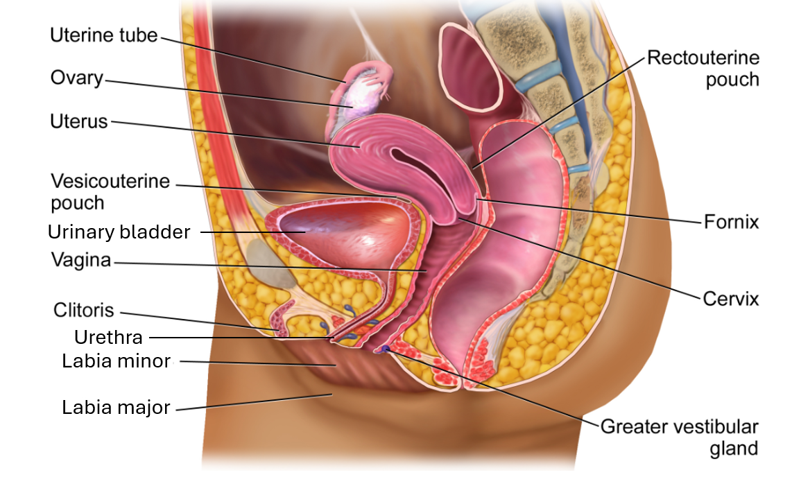 Figure 9.4: The female reproductive system
GONADS
The female gonads are the pair of ovaries. These are similar to the male testes in that they produce gametes and sex hormones. Ovaries produce ova or eggs, and female sex hormones estrogen and progesterone. The ovaries are located within the pelvic cavity, one on either side of the uterus.
The ovaries undergo a 28-day process called the ovarian cycle. The ovarian cycle has three phases: follicular phase, ovulation, and luteal phase. The follicular phase occurs between Day 1 and Day 14 of the cycle. It is when the follicles, fluid-filled sacs, enclose and nurture the eggs as they develop. The follicles release estrogen and a low level of progesterone, which indirectly helps the follicles mature. Ovulation is when the mature follicle ruptures and releases the egg into the uterine tube. This occurs around Day 14. The luteal phase occurs after ovulation from Day 15 to Day 28. The ruptured follicle transforms into the corpus luteum, which produces high levels of progesterone and lower levels of estrogen. These hormones help prepare the uterus for potential pregnancy. If the egg is not fertilized, then the corpus luteum degrades and the cycle repeats. (Figure 9.5)
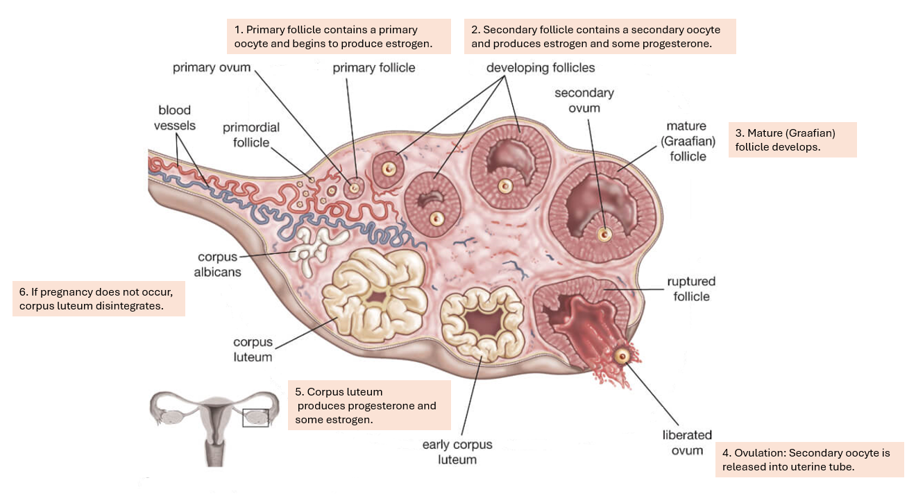 Figure 9.5: Ovarian Cycle
DUCT SYSTEM
After the ovum is released from the ovary during the ovarian cycle, it enters the uterine tube. The uterine tube is the site of fertilization if sperm are present. If the egg is fertilized, it travels through the uterine tube to the uterus. The uterus is a muscular, hollow, pear-shaped organ that has three layers: perimetrium (outermost layer), myometrium (thick middle muscular layer), and endometrium (inner lining). The fertilized egg (called a zygote) develops through cell division into the early embryo and will implant into the thickened endometrium. The uterus houses the embryo as it becomes a fetus. Eventually the fetus receives its nourishment through the placenta. If the egg is not fertilized, it will degrade and be shed during a female’s menstrual period along with the thickened endometrium that was built up in preparation for potential pregnancy.
The narrow inferior portion of the uterus is the cervix, which projects down into the vagina. The cervix produces mucus secretions that can block or allow sperm to enter the uterus. When estrogen levels are high, the secretions become thin and clear, helping sperm move through the female reproductive tract towards the egg. The upper portion of the vagina, or fornix, is a recess that surrounds the cervix. It acts as a reservoir for semen after ejaculation, allowing for seminal fluid to liquefy and move more easily through the cervix into the uterus.
VAGINA
The vagina is a muscular canal that serves as the copulatory organ in females. It is the structure for sexual intercourse, and also serves as the exit from the uterus during menstruation and childbirth. The vagina has an acidic environment that is maintained by the normal microbiome (a healthy community of bacteria, including predominantly Lactobacillus) that prevents potential pathogens from causing vaginal infection. Semen is slightly alkaline to help neutralize the vagina’s acidity, providing a suitable environment for the sperm.
EXTERNAL ORGANS: VULVA
The external female organs are collectively known as the vulva. The primary function of the vulva is to protect the internal reproductive organs and facilitate sexual activity. The labia majora and labia minora are two layers of skin folds that surround the clitoris, urethral opening, and vaginal opening. The clitoris is homologous to the male penis, with its focus being on sexual arousal. Within the labia and near the vaginal opening, the vestibular glands secrete a lubricating fluid, which helps provide vaginal and vulvar lubrication during sexual intercourse.
Activity 3: Identify the Function of the Organs of the Female Reproductive System
Match the female reproductive organ with its function. Record answers on the Lab Report.
|
Term |
Definition |
|
a. Vulva |
1. Female gonad |
|
b. Cervix |
2. Recess of vagina to collect semen |
|
c. Uterus |
3. Fluid-filled sac where eggs develop; produces estrogen |
|
d. Ovary |
4. Female gamete |
|
e. Uterine tube |
5. Collection of external female organs |
|
f. Clitoris |
6. Secretes mucus that helps sperm movement |
|
g. Vestibular glands |
7. Houses developing fetus |
|
h. Fornix |
8. Copulatory organ and birth canal |
|
i. Vagina |
9. Site of fertilization |
|
j. Follicle |
10. Primary site of sexual arousal |
|
k. Ovum |
11. Secretes a lubricating fluid for sexual intercourse |
Activity 4: Trace the Path of an Egg through the Female Reproductive System
Label female organs in the pathway of egg from ovulation to implantation to birth. Record answers on the Lab Report.
EMBRYONIC DEVELOPMENT
Embryonic development is divided into three stages: cellular, tissue, and organ development.
CELLULAR STAGE
Cellular development begins at fertilization. Fertilization occurs when a spermatozoan cell penetrates an egg and the nuclei of the haploid sperm and haploid egg fuse, creating a diploid zygote, which now contains a complete set of 46 chromosomes. As it travels through the uterine tube, the zygote then begins to divide through a process known as cleavage, where the single-celled zygote undergoes rapid mitotic division into smaller cells, doubling in number each time without growing larger. This cell division without growth continues until a cluster of 16-32 cells called a morula has been formed. By this point, 4 to 5 days after fertilization, the morula reaches the uterus, where it reorganizes into a blastocyst, a hollow ball of cells with a cluster of cells called the inner cell mass. The inner cell mass contains all the cells needed to produce the embryo whereas the outer layer of the blastocyst forms the placenta and other structures to sustain the pregnancy. During the second week of development, the blastocyst implants into the endometrium of the uterus. The endometrium grows over and surrounds the blastocyst, fully securing it to the uterine lining. The blastocyst begins to secrete the hormone human chorionic gonadotropic (hCG), which sustains early pregnancy and ensures the continued development of the placenta. (Figure 9.6)
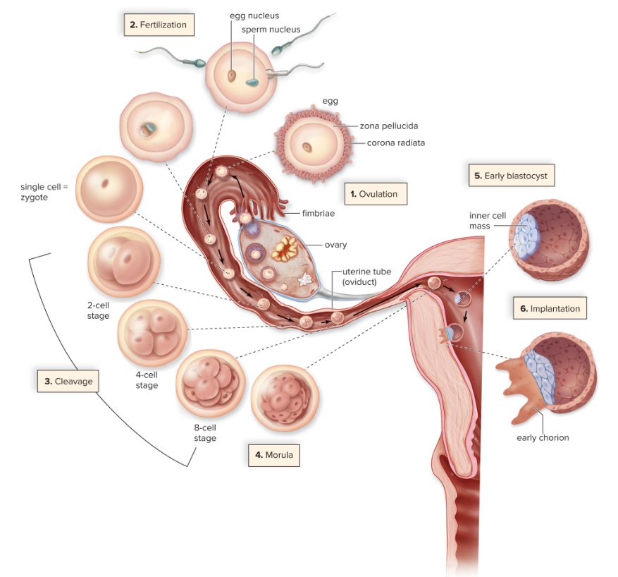
Figure 9.6: The cellular stage of embryonic development © McGraw Hill Education
Activity 5: View Cellular Stages of Embryonic Development under the Microscope
- Obtain a prepared slide of starfish development and view under the microscope (See Lab Exercise 1: How to Focus a Microscope).
- Record your observations of zygote, cleavage stage (2-cell stage, 4-cell stage, 8-cell stage, morula), and blastula stage.
TISSUE STAGE
During the third week of embryonic development, the tissue stage begins through the process of gastrulation. Gastrulation involves reorganizing the cells of the blastocyst (one-dimensional layer) into a three-dimensional, multi-layered structure. In early gastrulation, a row of cells folds inward, a process called invagination. This invagination produces a cavity known as the archenteron, which will form the gut cavity, and creates two distinct germ layers: ectoderm and endoderm. In late gastrulation, the third middle layer, mesoderm, is formed as a result of cell migration and interactions between the endoderm and ectoderm. (Figure 9.7)
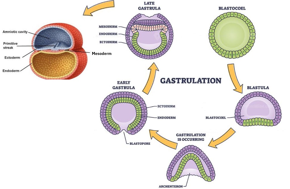
Figure 9.9: Gastrulation is when invagination of the blastocyst forms a cavity and eventually three germ layers.
ORGAN STAGE
Organogenesis is the final stage of embryonic development that starts at the end of gastrulation, beginning approximately in the fourth week and continuing through the eighth week. During organogenesis, the three germ layers differentiate into different organs and systems. The ectoderm is the outer layer and will give rise to the skin, nervous system and sensory organs. The mesoderm is the middle layer and will form connective tissues, muscles, bones, cardiovascular system, and internal organs, such as the kidneys and gonads. The endoderm is the inner layer and will develop the lining of the digestive and respiratory tracts as well as associated organs, such as the liver and pancreas. (Figure 9.10)
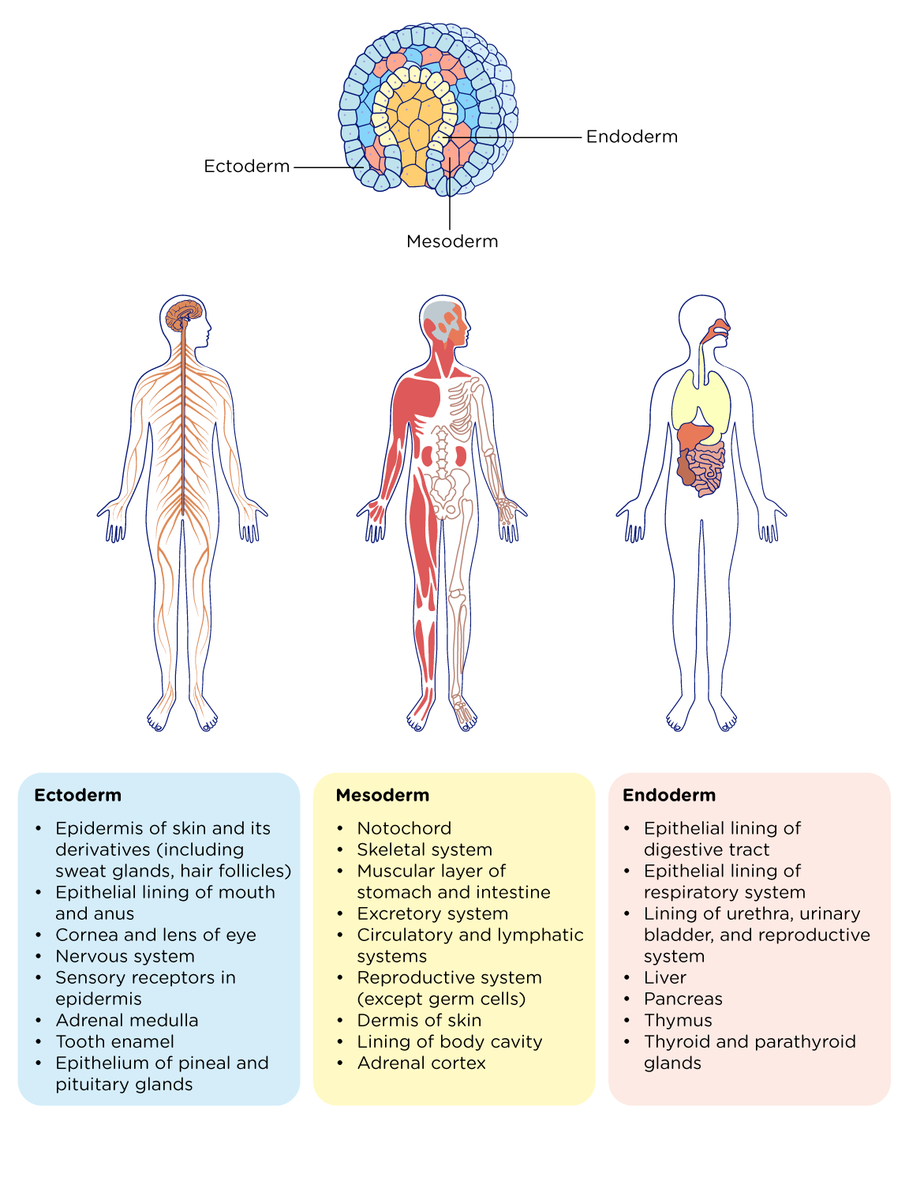 Figure 9.10: Organogenesis is the process of each germ layer differentiating into a specific organ and/ or system.
Figure 9.11 illustrates the external anatomy of a developing embryo between weeks 3 and 8 when organogenesis is occurring.
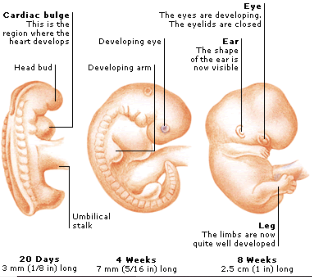
Figure 9.11: External anatomy of developing embryo
During embryonic development when major organs and body systems are forming, a baby is highly vulnerable to environmental factors that could cause birth defects. Examples are the mother exposing the developing embryo to drugs or alcohol; infections such as HIV, Zika virus, or rubella; poor nutrition; and various forms of radiation.
FETAL DEVELOPMENT
At around the ninth week, the embryo is considered a fetus, and fetal development continues from that point until birth. It is a period of rapid growth and development. The fetus develops all major organ systems, limbs and body parts. The skeleton develops from cartilage into bones, the reproductive organs form, and structures such as fingernails, eyelashes, and hair grow. Throughout this process the fetus enlarges in size and gains weight. (Figure 9.12) When it reaches full term, at about 40 weeks, the birth process occurs, producing a fully developed newborn baby.
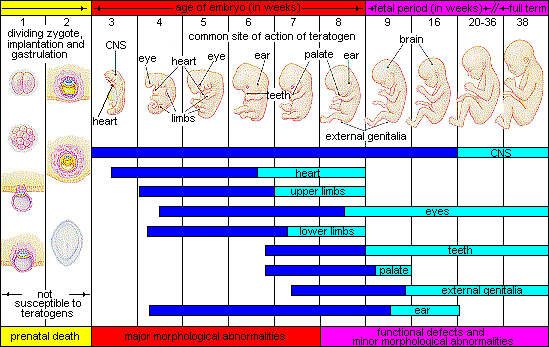
Figure 9.12: Timeline from Fertilization through the end of Fetal Development.
Activity 6: From Fertilization until the end of Fetal Development
Match the terminology with its definitions. Record your answers on the Lab Report.
|
Term |
Definition |
|
a. Blastocyst |
1. Middle layer of gastrula; gives rise to muscles, bones, and internal organs |
|
b. Gastrulation |
2. Last step of cleavage; 16 cells |
|
c. Cleavage |
3. Process of forming organs from the 3 germ layers |
|
d. Mesoderm |
4. Stage from week 9 until birth |
|
e. Fetal development |
5. A hollow ball of cells |
|
f. Fertilization |
6. Process in which the zygote undergoes a series of mitotic divisions |
|
g. Zygote |
7. Inner layer of gastrula; gives rise to lining of digestive & respiratory tracts as well as associated organs |
|
h. Endoderm |
8. Process of sperm fusing with egg |
|
i. Organogenesis |
9. Process of reorganizing cells of blastocyst into 3 germ layers |
|
j. Ectoderm |
10. A single, diploid cell that results from the fusion of sperm and egg |
|
k. Morula |
11. Outer layer of gastrula; gives rise to skin, nervous system & sense organs |
Activity 7: Lab Review
On the Lab Report, answer the questions in the Lab Review section.
Link to Lab Report: Lab 9 Reproduction & Development Lab Report
REFERENCES
Betts, J. G., Young, K. A., Wise, J. A., Johnson, E., Poe, B., Kruse, D. H., Korol, O., Johnson, J. E., Womble, M., & DeSaix, P. (2022d, April 20). 27 The Reproductive System – Anatomy and Physiology 2e | OpenStax. https://openstax.org/books/anatomy-and-physiology-2e/pages/27-1-anatomy-and-physiology-of-the-testicular-reproductive-system
AnatomyTOOL.org. (2014). Blausen 0400 – Female reproductive system (Lateral view) – English labels. Source: “Medical gallery of Blausen Medical 2014” https://en.wikiversity.org/wiki/WikiJournal_of_Medicine/Medical_gallery_of_Blausen_Medical_2014
Jack Westin MCAT Prep. (n.d.) Major Structures Arising Out of Primary Germ Layers. https://jackwestin.com/resources/mcat-content/embryogenesis/major-structures-arising-out-of-primary-germ-layers
Mader, Sylvia S. (2023). Laboratory Manual for Human Biology. 17th edition. McGraw-Hill.
New York-Presbyterian. (n.d.). Female Reproductive System | NYP. https://www.nyp.org/healthlibrary/multimedia/female-reproductive-system
Quizlet-Sarah Branning. (2018). Correctly Label the Following Parts of the Male Reproductive System. https://quizlet.com/296291669/correctly-label-the-following-parts-of-the-male-reproductive-system-diagram/?x=1jqt
Sokeane OBGYN. (n.d.). Information for new obstetrics patients. Ppt Download. https://slideplayer.com/slide/12566686/
Sapkota, A. (2023, August 3). Oogenesis / Ovulation / Ovarian cycle- Definition, Phages, Process. Microbe Notes. https://microbenotes.com/oogenesis-ovulation-ovarian-cycle/
Science Photo Gallery. (2025). Female Reproductive System #128. https://sciencephotogallery.com/featured/128-female-reproductive-system-pixologicstudioscience-photo-library.html
Shutterstock. (2023). Gastrulation Stages. https://www.shutterstock.com/image-vector/gastrulation-stages-early-embryo-development-process-2306778329
Tortora, Gerard J. and Bryan H. Derrickson. (2020). Principles of Anatomy and Physiology, 16th edition. John Wiley and Sons.
