6 Nervous System
Learning Objectives
- Distinguish between central nervous system and peripheral nervous system.
- Identify the structures of a neuron and describe their functions.
- Identify the regions of the brain and their structures.
- Identify the lobes of the cerebrum.
- State the general function of each lobe and structure of the brain.
- Identify the major structures of the sheep brain.
- Compare and contrast structural differences between the sheep brain and the human brain.
- Identify white matter and gray matter within a cross-section view of the spinal cord.
- Distinguish between cranial nerves and spinal nerves based on location.
- Describe the components of a reflex arc.
- Perform the knee-jerk reflex.
- Conduct reaction time experiments and compare undistracted reaction time to distracted reaction time for at least two different types of distractions.
NERVOUS SYSTEM
The nervous system can be separated into two major divisions: the central nervous system and the peripheral nervous system. The central nervous system consists of the brain and spinal cord whereas the peripheral nervous system includes all other nerves that extend throughout the body. (Figure 6.1)
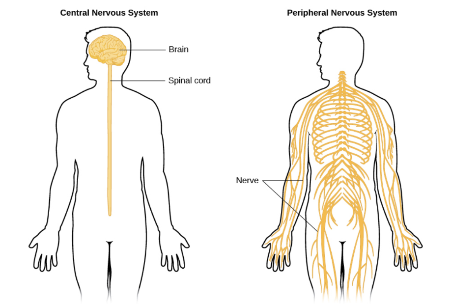 Figure 6.1: The nervous system is divided into two major parts: central nervous system and peripheral nervous system.
Nervous tissue is composed of two main types of cells: neurons and neuroglial cells. Neurons, or nerve cells, are the structural and functional units of the nervous system that receive, process, and transmit information through both electrical and chemical signals. They are made up of a cell body, or soma; dendrites; an axon; and synaptic knobs or buttons (Figure 6.2). The cell body can structurally be compared to the basic structure of an animal cell due to housing the nucleus and other essential organelles for the neuron’s function. The dendrites are branched extensions of the cell body that receive signals from other neurons. The axon is a long, cable-like projection that extends from the cell body of the neuron and transmits electrical signals to the other end of the neuron. The synaptic knobs or buttons are specialized ends of the axon that form a synapse with another neuron or cell. They transform the electrical signal into chemical signals, the neurotransmitters, and transmit the signals to receptors in neighboring cells.
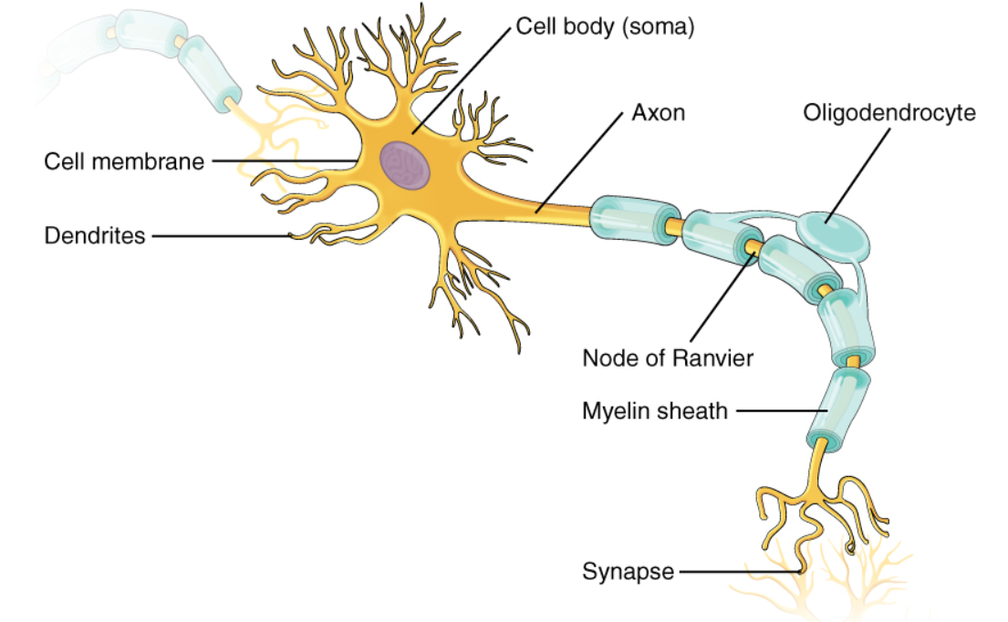
Figure 6.2: The basic structure of a neuron. © McGraw Hill Education
There are three main types of neurons: sensory neurons, interneurons, and motor neurons. Sensory neurons transmit electrical signals from sensory receptors in body tissues and organs to the central nervous system. They detect stimuli or sensations such as touch, heat, cold sound, pain, and light. Motor neurons transmit electrical signals from the central nervous system to muscles and glands and act in response to stimuli. They are involved in both voluntary and involuntary actions. Interneurons transmit electrical signals between sensory and motor neurons within the central nervous system.
Neuroglial cells, or glial cells, serve as support, protection, and insulation for neurons. Glial cells of the central nervous system include oligodendrocytes, astrocytes, ependymal cells, and microglial cells; glial cells of the peripheral nervous system include Schwann cells and satellite cells.
CENTRAL NERVOUS SYSTEM
BRAIN
The brain is composed of gray matter and white matter. Gray matter contains primarily the cell bodies of the neurons, and has its name based on appearing a grayish, pinkish color due to the presence of blood capillaries. White matter consists of the axons of the neurons which are covered in a white fatty structure called the myelin sheath. Gray matter is found mostly in the exterior and white matter is mostly in the interior of the brain.
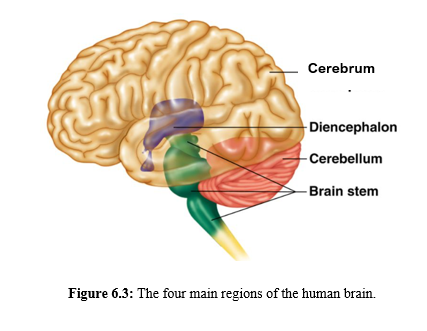
The brain has four main regions: the cerebrum, the diencephalon, the cerebellum, and the brainstem (Figure 6.3). Each of these regions can be further subdivided into multiple structures based on their functions.
 The cerebrum is the largest portion of the brain and is associated with higher mental function along with sensory and motor functions. It is divided into left and right cerebral hemispheres, joined at the corpus callosum. The surface area of the human brain is increased due to the folding of the brain tissue into gyri and sulci (folds and grooves), creating more surface area for more neurons to exist within the skull. The cerebrum is subdivided into lobes: frontal lobe, two parietal lobes, two temporal lobes, occipital lobe, and the insular lobe (Figure 6.4). The cerebral lobes contain distinct functional areas made up of sensory, motor, and association areas. The frontal lobe is primarily associated with motor functions (voluntary muscle control); cognitive functions, such as decision-making, problem-solving, and personality; and speech and language. The parietal lobes are associated with somatosensory reception, such as taste and tactile senses of touch, pressure, tickle, pain, and itch. The temporal lobes are associated with hearing and smell. The occipital lobe is responsible for visual perception. The insular lobe is hidden below the other lobes of the brain and plays a role in emotions and self-awareness.
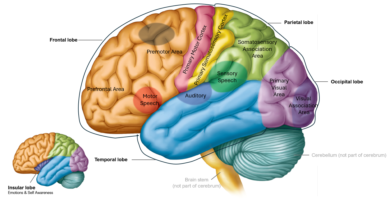
Figure 6.4: Five lobes of the cerebrum along with their functional areas © McGraw Hill Education
Note to students: Write all data and answers to questions on the Lab Report provided.
Activity 1: Match the Functions to the Cerebral Lobes
Match the cerebral lobe with its function. Record your answers on the Lab Report.
|
Lobe |
Definition |
|
a. Frontal |
1. Vision perception |
|
b. Parietal |
2. Hearing and smell |
|
c. Temporal |
3. Cognitive function; personality; motor function |
|
d. Occipital |
4. Emotions and self-awareness |
|
e. Insular |
5. Somatosensory reception (touch and taste) |
The diencephalon connects the cerebrum with the rest of the nervous system except for the sense of smell. The olfactory tract has a direct connection from the olfactory epithelium, found at the top of the sinuses, to the cerebrum. The diencephalon includes the thalamus, hypothalamus, epithalamus, and subthalamus. All sensory information, except for the sense of smell, passes through the thalamus before reaching the cerebrum. The thalamus can be considered the Grand Central Station of senses as it also processes that sensory information. The other major region of the diencephalon is the hypothalamus, which is composed of a collection of nuclei that serves as one of the major regulators of homeostasis. It contains control centers for body temperature, blood pressure and heart rate, water and electrolyte balance, appetite and weight control, and the sleep-wake cycle. The epithalamus, which contains the pineal gland, also regulates the sleep-wake cycle by secreting the hormone melatonin. The subthalamus regulates motor activity and has a key role in controlling voluntary movement.
Activity 2: Match the Functions to the Structures of the Diencephalon
Match the structure of the diencephalon with its function. Record your answers on the Lab Report.
|
Structure |
Definition |
|
a. Hypothalamus |
1. Regulates sleep-wake cycle through the hormone melatonin |
|
b. Epithalamus |
2. Regulates motor activity and controls voluntary movement |
|
c. Subthalamus |
3. Processes all sensory information except the sense of smell |
|
d. Thalamus |
4. Regulates homeostasis |
The cerebellum is located posterior to the cerebrum. It has the function of coordinating and fine-tuning voluntary muscle movements; helping to maintain balance and equilibrium; and plays a role in motor learning, such as walking and riding a bicycle.
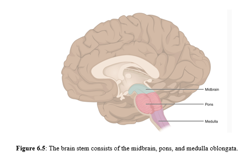
The brain stem is the part of the brain that connects it with the spinal cord. It serves as a relay station for nerve impulses passing from the spinal cord to the brain. It consists of 3 components: midbrain, pons, and medulla oblongata (Figure 6.5). The midbrain plays an important role in processing visual, auditory, and somatosensory perception. The pons connects the brain stem to the cerebellum and acts as a bridge to relay nerve impulses between the two structures. The medulla oblongata regulates breathing, heart rate, and blood pressure. It also plays a role in reflexes such as swallowing, vomiting, coughing, and sneezing. It is the main connection between the brain and the spinal cord.
Activity 3: Match the Functions to the Structures of the Brain Stem
Match the structure of the brain stem with its function. Record your answers on the Lab Report.
|
Structure |
Definition |
|
a. Midbrain |
1. Serves as the control center for breathing, heart rate, and blood pressure |
|
b. Pons |
2. Connects the brain to the cerebellum to relay the nerve impulses |
|
c. Medulla oblongata |
3. Processes visual, auditory, and somatosensory perception |
Activity 4: Dissection of the Sheep Brain
The sheep brain is similar to the human brain as both organisms are mammals. However, some regions of the sheep brain are proportionally smaller compared to the human brain. For example, the sheep cerebrum is considerably smaller in proportion to total brain size than the human cerebrum. The orientation of the brain stem and spinal cord is also different because humans are bipedal (walk upright on two legs) and sheep are quadrupedal (walk on four legs). The sheep brain stem and spinal cord emerge and run horizontally from the back of the brain whereas the human brain stem and spinal cord emerge and run vertically below the cerebrum.
EXTERNAL BRAIN ANATOMY
- One student from each lab bench may obtain safety glasses, a pair of nitrile gloves, one preserved sheep brain, one scalpel, one pair of scissors, one probe, and one dissecting tray.
- Remove the sheep brain from its packaging and throw away the packaging in the regular garbage near the lab bench. Place the sheep brain dorsal side (top side) up onto the dissecting tray.
- Observe the external anatomy:
- The brain and spinal cord are enclosed in three membrane layers called the meninges that serve as protection. These meninges, from outermost to innermost, are the dura mater, arachnoid mater, and pia mater. The dura mater is the tough, thick, cloudy outer layer. The arachnoid mater and pia mater form a thin covering that adheres to the surface of the cerebrum.
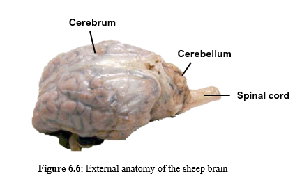
- Using Figure 6.6, identify the cerebrum, cerebellum, and spinal cord.
- Remove the dura mater by cutting it with scissors and gently pulling it from the surface of the brain.
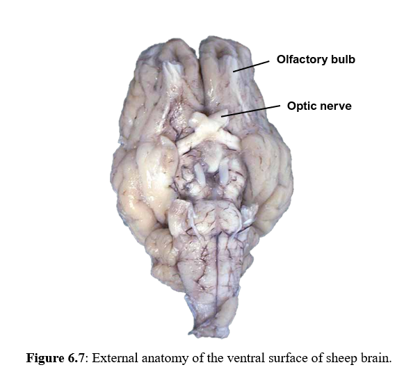
- Position the brain with its ventral (bottom) surface up. Using Figure 6.7, identify the olfactory bulbs and optic nerve.
INTERNAL BRAIN ANATOMY
- Return the sheep brain to the dissecting tray, dorsal surface up.
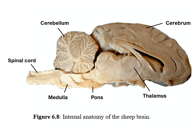
- Using a scalpel, cut the sheep brain along the midline through the cerebrum, cerebellum, and spinal cord to separate the brain into 2 longitudinal halves.
- Observe the internal anatomy: Using Figure 6.8, identify the cerebrum, cerebellum, spinal cord, thalamus, pons, and medulla oblongata.
- Cut a cross section of the frontal lobe to view white matter and gray matter. (Figure 6.9)
- After all observations have been carried out, properly dispose of the dissected sheep brain by placing it in the designated pail near the instructor table.
- Wash all dissecting tools and return to the tray of tools. Wash the dissecting tray and return to the stack of dissecting trays on the side lab bench. Take off your gloves and wash your hands with soap.

Record the answers to the following questions on the Lab Report:
Compare the sheep brain to the human brain:
- Are there any proportional differences in the size of these brain regions: cerebrum, cerebellum, or brain stem? If so, what are they?
- Are there any positional differences of these brain regions: cerebrum, cerebellum, or brain stem? If so, what are they?
- What are the differences in the number and depth of the gyri (folds) and sulci (grooves) of the cerebrum?
- What is the difference in the size of the olfactory bulbs between sheep and human brains? Why would this size difference occur?
SPINAL CORD
The spinal cord is also composed of gray and white matter that is distributed differently compared to the brain. In the spinal cord, gray matter is the most interior, forming a butterfly-shape, and white matter is the most exterior (Figure 6.10). The spinal cord acts as the main communication pathway between the brain and the rest of the body as it begins at the end of the brain stem and extends down through the vertebral column of the back, ending as the conus medullaris in the lower back, between the first and second lumbar vertebrae.

Figure 6.10: A cross section of the spinal cord, showing gray matter in a butterfly shape surrounded by white matter.
PERIPHERAL NERVOUS SYSTEM
The peripheral nervous system contains cranial nerves and spinal nerves. Twelve pairs of cranial nerves project from the ventral (bottom) surface of the brain, connecting it to various parts of the head, neck, and upper trunk. These nerves transmit information that controls functions like vision, smell, taste, hearing and facial movement. Thirty-one (31) pairs of spinal nerves emerge from the spinal cord, transmitting both sensory and motor signals between the central nervous system and the rest of the body. These nerves are vital for transmitting information related to movement, sensation, and autonomic functions.
REFLEXES
Spinal nerves play a role in spinal reflexes, which are rapid, involuntary responses to stimuli. These reflexes occur through a reflex arc involving sensory nerves, spinal cord interneurons, and motor neurons, without communicating with the brain (Figure 6.11). An example of a spinal reflex is the withdrawal reflex that involves heat and pain. You instinctively pull your hand away from a hot surface or sharp object prior to consciously feeling the pain.

Figure 6.11: Spinal Reflex (specifically Withdrawal Reflex): the arrow illustrates the path of the nerve impulse traveling from a sensory receptor to motor effector.
Activity 5: Performing a Spinal Reflex
Another example of spinal reflex is a stretch reflex where stretch receptors of a tendon detect a stimulus, such as a tap and send the sensory nerve impulse to the interneurons in the spinal cord. Then the interneurons transmit to the motor neurons, which transmit to the effector, in this case, a muscle, causing muscle contraction and a jerking motion results. One example of a stretch reflex is the knee-jerk reflex (patellar reflex) where the patellar tendon is tapped with a reflex hammer and the quadriceps femoris muscle contracts, causing the lower leg to jerk outward.
Work in pairs to carry out this lab activity:
- Have the subject sit on a lab chair or the lab bench, so that their legs hang freely.
- Using the reflex hammer sharply tap one of the patellar tendons just below the kneecap.
- Observe and record your results on the Lab Report.
- Repeat steps 1-3 for the other leg.
- Switch roles and repeat steps 1-4.
REACTIONS
A reaction is a voluntary response to the reception of a stimulus. Voluntary means that your conscious mind initiates the reaction. An example is swatting a fly once it has landed in an accessible spot. Because neurons must carry the sensory message to the cerebral cortex and the motor neuron to react, a reaction takes more time than a reflex. Reaction time has the following components:
- The time it takes for the stimulus to reach the receptor unit.
- The time it takes for the receptor to process the message.
- The time it takes for a sensory neuron to carry the message to the integration center.
- The time it takes for the integration center to process the information.
- The time it takes for a motor neuron to carry the response to the effector.
- The time it takes for the effector to respond.
This exercise involves measuring reaction time under various conditions. One student will serve as the investigator conducting the experiment, while another student is the subject. Then switch roles so that each student has the opportunity to serve as investigator or subject.
Visual reaction time can easily be measured with a reaction-time stick. This device makes use of the principle of progressive acceleration of a falling object. The acceleration of Gravity (g) is 9.8 m/s/s. That means that in freefall, at 1 second after the release the object’s speed is 9.8 meters/second. But this speed increases by 9.8 meters per second, so that by two seconds the object is moving twice as fast, at 19.6 meters per second. How fast is it moving at 3 seconds?
You can see from the Reaction Time stick that the distance the stick drops increases the longer it takes to catch it. (The “time” intervals get longer as you go up the stick.)
How fast can you catch the Reaction Time stick? The measurements are in milliseconds (thousandths of a second) and the labels range from 50 msec to 400 msec. Each “test” will take no more than half a second.
Your reaction to the dropped stick is being measured. Your nervous system must sense and respond to the stimulus: a dropped stick. Does your environment affect the way your nervous system works? Will you have different reaction times under different conditions? Follow the directions to design and conduct the experiments.
Activity 6: Measuring Reaction Time
Work in pairs:
Decide what kind of distraction you will be testing. Set up the conditions for undistracted and distracted testing. Some examples of distractions are included below. Once you have decided on the distraction, be consistent about it through the 10 “distracted” tests.
Choose (and describe) what distraction you are testing on the Lab Report:
___ Facebook
___ Instagram
___Watching a video (what video – website?)____________________
___Playing Candy Crush or other game (name of game?)______________
___Texting a friend
___Reading email
___Writing email
___Listening to music
___Other distraction (explain)_______________________________
You will complete two sets of 10 tests: one set of undistracted tests and the other set with distracted conditions. You must alternate undistracted with distracted tests until you complete 10 of both.
Why would you design the experiment with alternating the type of tests rather than just doing all ten of one condition followed by all ten of the other condition?
- Have the subject sit on a lab chair.
- The investigator stands facing the subject and holds the stick in a vertical position; the release end of the reaction-time stick is held with the thumb and forefinger of the dominant hand, at eye level or higher.
- The subject positions their thumb and forefinger of the dominant hand around the thumb line near the bottom of the stick. The space between the subject’s thumb and forefinger should be about 1 inch.
- The subject tells the investigator when they are ready to be tested.
- Once the investigator is told the subject is ready, at any time during the next 10 seconds, the investigator releases the reaction time stick.
- The subject catches the ruler between the thumb and forefinger as soon they can after as it starts to fall.
- After it is caught, the line under their thumb represents visual reaction time in milliseconds.
- The subject reads the reaction time from the ruler out loud, and the investigator records the data in Table 1 on the Lab Report.
- Repeat steps 1 through 7 until you have (alternating) 10 of undistracted and 10 of distracted conditions recorded. Calculate the average reaction time from each set of 10 trials.
- Repeat steps 1 through 8 for each member of the group.
- Repeat the experiment, undistracted and distracted (steps 1 through 9) with a different distraction and record data in Table 2 on the Lab Report.
Within each trial the reaction times of most of the ten trials should be similar, but perhaps the first few (or one at random) may be relatively different from the others. Suggest some reasons for this variability. Record on the Lab Report.
12. The instructor may collect the data from your experiments by having each student post their average reaction times (undistracted and distracted) and the types of distractions on the board. Compare your results with other students. How do different distractions affect reaction time? Why might different people have different reaction times?
Activity 7: Lab Review
On the Lab Report, answer the questions in the Lab Review section.
Link to Lab Report: Lab 6 Nervous System Lab Report
REFERENCES
Berry, Miles. (modified 2017). The Brain Ch. 7c The Brain Functional Anatomy –Cerebral Hemispheres –Diencephalon –Brain Stem –Cerebellum. – ppt download. https://slideplayer.com/slide/10991084/
Betts, J. G., Young, K. A., Wise, J. A., Johnson, E., Poe, B., Kruse, D. H., Korol, O., Johnson, J. E., Womble, M., & DeSaix, P. (2022, April 20). Anatomy and Physiology 2E | OpenStax. https://openstax.org/books/anatomy-and-physiology-2e/pages/12-2-nervous-tissue#fig-ch12_02_01
Carolina Biological. (n.d.) Teacher Resources- Sheep Brain Dissection. https://www.carolina.com/teacher-resources/Interactive/sheep-brain-dissection/tr10991.tr
Carolina Biological. (n.d.) Reaction Time Experiment.
Libretexts. (2021, August 1). 11.7: Sheep brain dissection. Biology LibreTexts. https://bio.libretexts.org/Bookshelves/Human_Biology/Human_Anatomy_Lab/11%3A_The_Central_Nervous_System_(Brain)/11.07%3A_Sheep_Brain_Dissection
Libretexts. (2022, August 9). 4.1.2: Parts of the Nervous System. Social Sci LibreTexts. https://socialsci.libretexts.org/Courses/Heritage_University/Introductory_Psychology_-_Heritage_University/04%3A_Week_4_-_Biological_Psychology/4.01%3A_Class_Day_4.1/4.1.02%3A_Parts_of_the_Nervous_System#:~:text=parasympathetic%20nervous%20system.-,Summary,from%20the%20central%20nervous%20system.
Mader, Sylvia S. (2023). Laboratory Manual for Human Biology. 17th edition. McGraw-Hill.
Mukherjee, S. (2023, April 29). Reflex Arc – Definition, steps, components, and diagram. Science Facts. https://www.sciencefacts.net/reflex-arc.html
Snider, Phillip and Terry Martin. (2024). Laboratory Manual to accompany Hole’s Essentials of Human Anatomy and Physiology. McGraw-Hill Publishing.
Tortora, Gerard J. and Bryan H. Derrickson. (2016). Principles of Anatomy and Physiology, 15th edition. John Wiley and Sons.
Wahrman, Miryam. (2019). Reaction Time Experiment (modification). Wayne, NJ: William Paterson University.
