3 Mammalian Dissection
Learning Objectives
- Identify the external anatomy of a fetal pig.
- Determine the sex of a fetal pig based on external anatomy.
- Identify and locate major organs and structures of the cardiovascular, respiratory, urogenital and digestive systems within the fetal pig.
- State the functions of the major organs and structures of the cardiovascular, respiratory, urogenital and digestive systems of the fetal pig.
- Compare and contrast fetal pig anatomy with human anatomy.
Dissecting a fetal pig allows for three-dimensional observations of the structure of a mammal that is comparable to a human, viewing the interconnections between organs and their systems. The fetal pigs that are available for dissection have been double injected with latex. Red latex has been injected into the arterial system whereas blue latex has been injected into the venous system.
EXTERNAL ANATOMY
Fetal pigs and humans are examples of mammals, characterized by being warm-blooded, having hair or fur, having a four chambered heart, and having mammary glands to produce milk for their young. Pigs and humans are also both placental mammals, where embryonic and fetal development occurs within the uterus of the mother. The developing young’s umbilical cord is connected to the mother via the placenta where nutrients, wastes, and gases are exchanged. After birth, the young mammals are able to receive nourishment by suckling on the mother’s lactating nipples of their mammary glands.
Note to students: Write all data and answers to questions on the Lab Report provided.
Activity 1: Examine the External Anatomy of the Fetal Pig
Working in groups of 2 to 4:
- Collect one dissecting tray, one scalpel, one pair of dissecting scissors, one probe, two long pieces of string, one pair of gloves per student, one pair of safety glasses per student, and one fetal pig. Put on safety glasses and gloves before starting the dissection.
- While standing near a lab sink, cut open the bag of the fetal pig and drain all fluid down the drain while the faucet is turned on. (Please note this liquid is a preservative solution that is a formaldehyde-free alternative. It is a propylene glycol-based solution called BioShield and is safe to work with in the lab and dispose of down the sink.)
- Position the fetal pig on its side on the dissecting tray. Locate the following structures of its external anatomy by comparing to Figure 3.1:
- External ear
- Eyelid
- Mouth
- Nose
- Nipples
- Umbilical cord
- Digits
- Tail
- Anus (under the tail)
4. Become familiar with the following anatomical directional terms:
- Dorsal: toward the back
- Ventral: toward the belly
- Anterior: toward the head
- Posterior: toward the rear or hind region
- Midline: vertical line that divides the body into left and right halves
- Lateral: away from the midline of the body
- Medial: toward the midline of the body
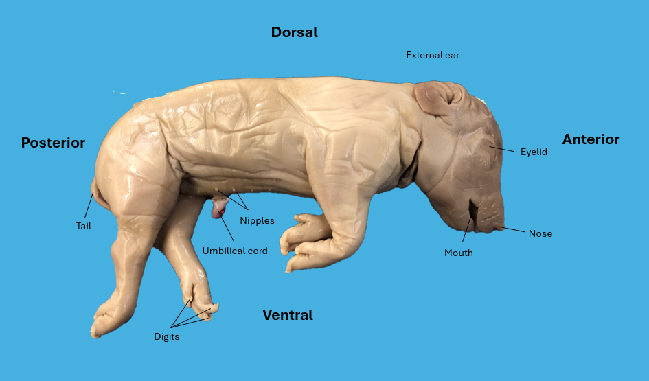
Figure 3.1: External anatomy of a fetal pig.
5. Determine the sex of the fetal pig by comparing to Figure 3.2:
- Female: Locate the urogenital papilla, the fleshy projection between the hind legs, on the ventral side. Locate the urogenital opening underneath the urogenital papilla.
- Male: Locate the urogenital opening just posterior to the umbilical cord on the ventral side. Just under the skin, locate the penis by feeling for a long, thick tube. Locate the scrotum just anterior to the anus and between the hind legs on the ventral side. The scrotum contains the testes, the male sex glands.
6. View a fetal pig of the opposite sex that another group is dissecting.
(a) 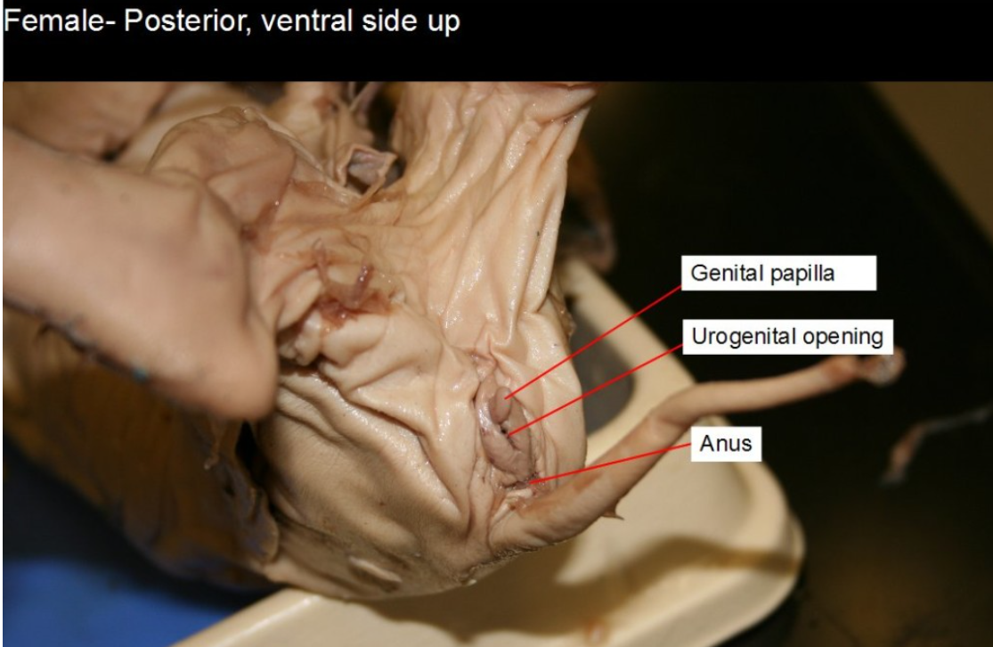
(b)
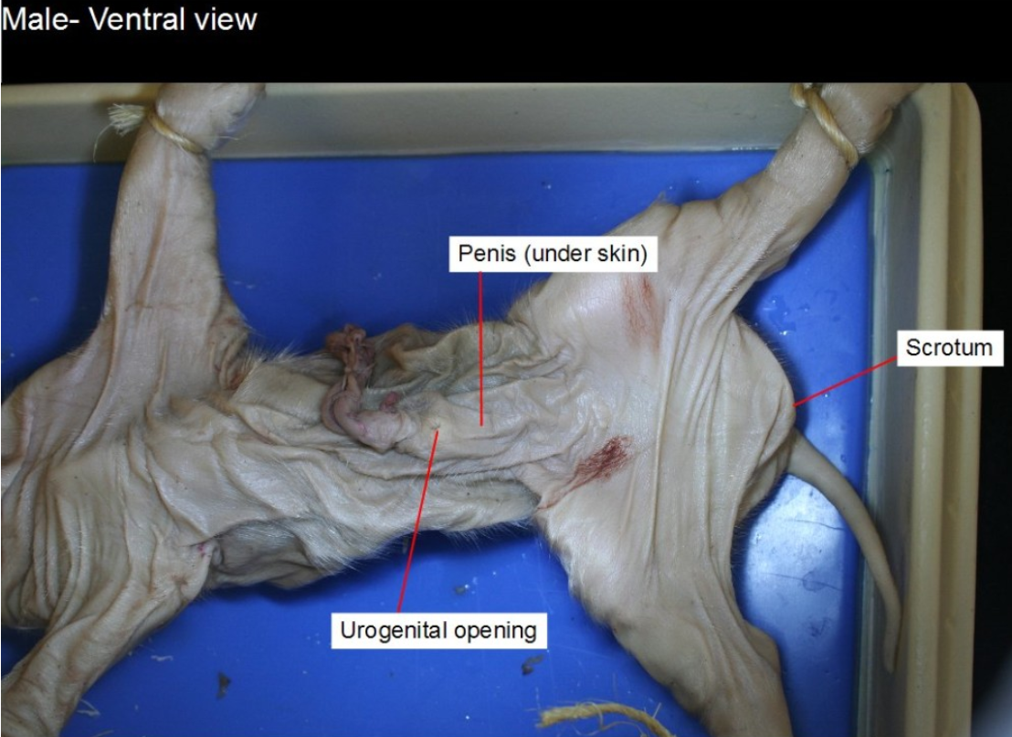
Figure 3.2: (a) Female anatomy of fetal pig; (b) Male anatomy of fetal pig. Note: Urogenital opening for both female and male fetal pigs is an opening for both the urinary and reproductive systems.
Answer the following questions on the Lab Report:
- Is the eyelid anterior or posterior to the umbilical cord?
- Is the umbilical cord anterior or posterior to the urogenital opening of the male fetal pig?
- Did you observe any hair or fur on the fetal pig? If so, where?
- Do male or female fetal pigs, or both, have nipples?
- What is the sex of your fetal pig?
INTERNAL ANATOMY
ORAL CAVITY AND PHARYNX
The oral cavity is also known as the mouth and contains the tongue, teeth, and salivary glands. The pharynx is just past the oral cavity dorsally and is also known as the throat. The pharynx is divided into three components: nasopharynx, oropharynx, and laryngopharynx. These structures are part of both the digestive and respiratory systems. At the laryngopharynx, the most inferior section of the pharynx, the digestive and respiratory systems separate, or diverge into the esophagus and trachea. The glottis is the opening that leads to the trachea, or windpipe, and lungs of the respiratory system. A flap of cartilage called the epiglottis covers the glottis when food is being swallowed, to keep food from entering the trachea. When the epiglottis covers the glottis, food is sent down the esophagus, the muscular food tube that is in the thoracic (chest) cavity and connects to the stomach of the digestive system.
Activity 2: Examine the Oral Cavity of the Fetal Pig
- Insert a pair of scissors into one corner of the fetal pig’s mouth and cut downward about 1.5 inches. Repeat on the opposite side.
- Place your thumb on the tongue and gently push downward on the lower jaw. This will widen the opening of the oral cavity to keep it open for observation. Be careful not to press on the teeth, as they can be sharp.
- Locate the following structures of the oral cavity:
- Tongue — fleshy, muscle that aids in chewing, swallowing, taste and speech.
- Underdeveloped teeth – may be sharp along the gum line, used for chewing.
- Hard palate – anterior bony portion of the roof of the mouth.
- Soft palate – posterior portion of the roof of the mouth, made up of soft tissue. Note: pigs do not have the uvula—extension of soft palate that hangs down in the back of the throat of humans. It prevents food and liquid from entering nasopharynx during swallowing.
- Epiglottis – flap of cartilage that covers the glottis during swallowing.
Answer the following questions on the Lab Report:
- What are the three portions of the pharynx?
- What is the common name for the pharynx?
- What structure prevents food and liquid from entering the trachea during swallowing?
- What structure prevents food and liquid from entering the nasopharynx during swallowing?
Activity 3: Dissection of Neck Region, Thoracic and Abdominal Cavities of the Fetal Pig
- Place the fetal pig ventral side up on the dissecting tray. Stretch the limbs so the pig is spread out on the tray.
- Tie one of the long strings around one of the forelimbs of the fetal pig, and then bring the string under the dissecting tray to the opposite side of the tray to tie the string to the other forelimb.
- Follow Step 2 for the hindlimbs.
- Using a scalpel, carefully create incisions according to the incision pattern seen in Figure 3.3. Incision #1 begins at the chin of the fetal pig. Use the scissors to assist in making the incisions, cutting horizontally to open the skin. Note: The dotted line between Incision #3 and #6 is not to be cut. This is to prevent damage to the diaphragm, the muscle that divides the thoracic and abdominal cavities. The diaphragm is one of the muscles that is responsible for breathing, particularly inhalation.
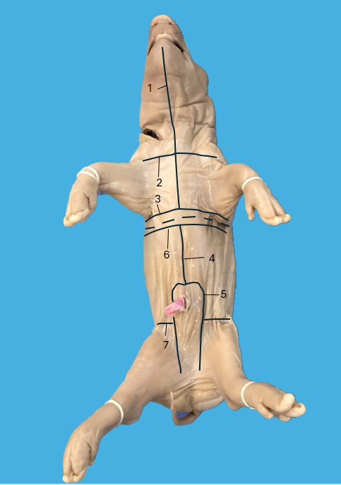
Figure 3.3: Numbered incision pattern for the dissection of the fetal pig. - After the thoracic and abdominal cavities have been properly dissected open, you may use paper towels to blot any excess fluid.
- There may be excess red or blue latex within the abdominal cavity. You can use the distilled water bottle to rinse the cavity of the latex and then blot with paper towels to remove any excess fluid.
- You may use dissecting pins to pin the flaps of the thoracic and abdominal cavities to the dissecting tray.
NECK REGION
- Separate the skin flaps from the neck region of the fetal pig more thoroughly with a scalpel or scissors.
- Locate the larynx, or voice box, in the upper neck region. It is a large, hollow structure that is composed of cartilage and contains the vocal cords. (Figure 3.4)
- Locate the trachea, or windpipe, which is the cartilaginous tube that extends in a posterior direction from the larynx and travels down behind the heart. The trachea is called the windpipe because air that is inhaled through the nose, goes down the pharynx, through the larynx, and into the trachea to reach the lungs for respiratory gas exchange to occur. The trachea has thin bands of cartilage along its length that hold the structure open for airflow.
- Use a probe or scalpel to move the trachea to the side to locate the esophagus behind it. The esophagus is a portion of the digestive tract that transports food from the pharynx to the stomach. It is a stretchy muscular tube that moves the food through contractions called peristalsis. (Figure 3.4)
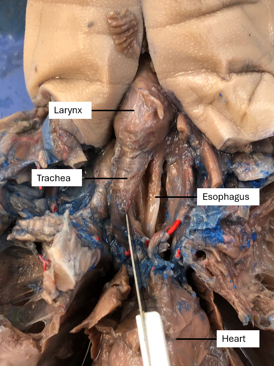 Figure 3.4: Neck region of a dissected fetal pig.
Answer the following questions on the Lab Report:
- What structure contains the vocal cords?
- What structure is also known as the windpipe?
- What structure is behind the trachea and is part of the digestive system?
THORACIC CAVITY
The thoracic cavity, also known as the chest cavity, is the space within the rib cage and above the diaphragm that houses the heart and lungs. The heart serves as the main pump for the cardiovascular system. It transports nutrients and oxygen to all the body tissues and organs and transports waste and carbon dioxide to the proper site of removal. The lungs are part of the respiratory system and are the site for gas exchange: bringing oxygen into the body and removing carbon dioxide from the body.
- Locate the heart within the thoracic cavity. The heart is contained within a pericardial sac, or loose membrane. Use a pair of scissors to open the pericardial sac and then pull it away from the heart.
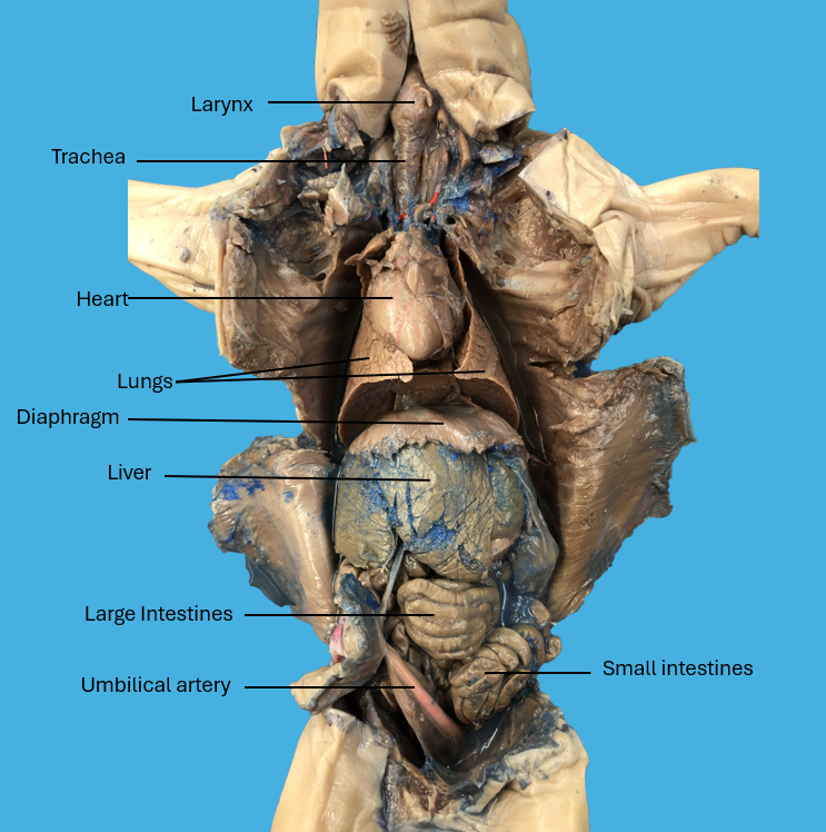
Figure 3.5: The major organs of the thoracic and abdominal cavities of a dissected fetal pig. - Examine the lungs on either side of the heart. Note that the right lung of the fetal pig has four lobes and the left lung has three lobes. The right lung of humans has three lobes and the left lung has 2 lobes.
- Compare the organs of the thoracic cavity of the fetal pig and humans by referring to Figure 3.5 and Figure 3.6 and the available models.
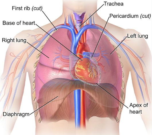
Figure 3.6: Thoracic cavity of the Human
Answer the following questions on the Lab Report:
- What structure divides the thoracic cavity from the abdominal cavity?
- What structure serves as the pump for the cardiovascular system?
- How do the lungs of the fetal pig differ from that of the human?
ABDOMINAL CAVITY
The abdominal cavity is a large, hollow space in the body that is located between the diaphragm and pelvic cavity, the space between the hip bones. It contains the liver, gall bladder, stomach, spleen, pancreas, small intestine, large intestine and kidneys. These organs are contained within the abdominal cavity by connective tissue called the greater omentum.
Refer to Figure 3.7 and Figure 3.8 to locate the abdominal organs in the fetal pig and compare to the human body.
- Locate the liver, a large, brown organ. It is the largest organ in the abdomen. The liver has numerous functions: 1) metabolism of carbohydrates, lipids, amino acids, nucleic acids, vitamins and minerals; 2) synthesis of blood proteins; 3) secretion of bile to aid in digestion; 4) detoxification of blood; 5) maintaining blood glucose levels by secreting the hormone glycogen; and 6) disposing of old red blood cells and recycling their iron.
- Locate the gall bladder by lifting the liver up and viewing the underside of the liver. The gall bladder stores the bile that is produced by the liver. It transfers the bile via the common bile duct to the small intestines, specifically the duodenum, the first segment of the small intestine, to assist in digestion of fats.
- Locate the following organs of the digestive system:
- Stomach is a large pink sac on the left side of the fetal pig’s abdominal cavity. You may need to move the liver to locate the stomach. The stomach stores food and releases gastric juices to digest proteins within the food. The stomach of a fetal pig is empty, as a fetal pig has never eaten.
- Small intestine is connected to the right part of the stomach in the back of the abdominal cavity. The first segment of the small intestines is the duodenum. The next two segments, jejunum and ileum, make up the long, irregularly coiled remainder of the small intestine that are held together by a common mesentery. The function of the small intestine as a part of the digestive tract is digest (chemically break down food) and absorb the nutrients and other products of food digestion.
- Large intestine (colon) is the other darker, thicker coiled bundle of tubes towards the right side of the abdominal cavity. At the junction of the ileum segment of the small intestines and the colon, there is a blind pouch called the cecum. The cecum houses bacteria that digest plant materials in the food that has been ingested. The layout of the colon of the fetal pig differs from that of humans. The colon of the fetal pig is coiled into a spiral shape, while the colon of a human makes a big loop around the small intestine in the abdominal cavity.
4. Locate the spleen by moving the stomach towards the center of the abdominal cavity. The spleen is a long, flat, reddish organ. It is part of the lymphatic system and serves as a filter for blood, removing damaged red and white blood cells. It is also the site of hematopoiesis, blood cell production.
5. Locate the pancreas by moving the liver and stomach up towards the diaphragm. It is a flattened, elongated organ with a spongy appearance. The pancreas is both an exocrine and endocrine gland. As an exocrine gland, it produces digestive enzymes to digest carbohydrates, fats and proteins and sodium bicarbonate to neutralize the acidity of the digested food of the stomach. As an endocrine gland, it produces the hormone insulin to lower blood glucose levels and the hormone glucagon to increase blood glucose levels.
6. Shift the intestines towards one side of the abdominal cavity to expose the kidneys. The kidneys are attached to the dorsal side of the cavity. Remove the connective tissue to make the kidneys more visible. Cut one of the kidneys longitudinally, dividing the kidney front to back, to view the interior. The kidneys serve as filters of the blood to remove waste products and excess fluid from the blood, producing urine. They regulate the body’s fluid and electrolyte balance.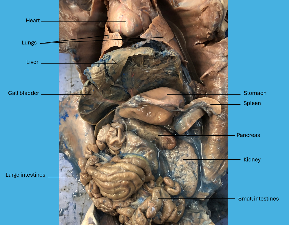
Figure 3.7: A closer look at the major organs of the abdominal cavity of a dissected fetal pig
(a)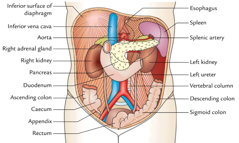
(b)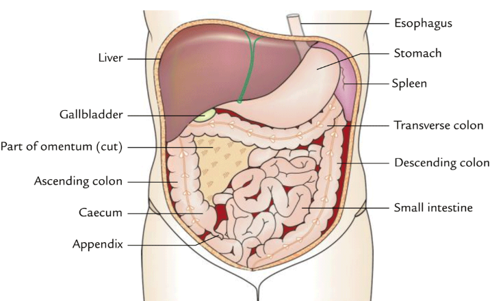
Figure 3.8: (a) Major organs in the dorsal section of the abdominal cavity of the human; (b) major organs in the ventral section of the abdominal cavity of the human.
Answer the following questions on the Lab Report:
- List the organs of the digestive system that are in the abdominal cavity.
- What organs are considered accessory organs to the digestive system?
- How does the large intestine of the fetal pig differ from that of the human?
- What system do the kidneys belong to?
PELVIC CAVITY
Posterior to the abdominal cavity you will find the pelvic cavity. This is where organs of the reproductive system are housed. If you are dissecting a male pig you can dissect the two testes by cutting the scrotal sacs open with a scalpel and using a probe or forceps to expose the testes.
If you are dissecting a female pig, it is possible to see two ovaries, small oval structures that produce eggs, and two slender coiled oviducts or uterine tubes that will conduct and hold the eggs for fertilization in a mature pig.
View the reproductive organs of a pig of the opposite sex that another group is dissecting.
PROPER CLEAN-UP PROCEDURE
- Place your fetal pig and all dissected components into its original bag and put it into the designated bucket as directed by your lab instructor.
- Wash and return all dissecting tools to the tray in the proper locations: dissecting pins, scalpels, scissors, and probes.
- Wash and return the dissecting tray to the stack of clean dissecting trays as directed by your lab instructor.
- Place all used gloves and paper towels in the regular garbage can.
- Return your safety glasses to the labeled drawer.
- Wash your hands.
Activity 4: Lab Review
On the Lab Report, answer the questions in the Lab Review section.
Link to Lab Report: Lab 3 Mammalian Dissection Lab Report
REFERENCES
Bio Corporation. (2016). Bio Corporation Safety Data Sheet. In Bio Shield Humectant Spray. https://cdn.shopify.com/s/files/1/0565/1611/6561/files/SDS_Bio_Shield_Humectant_Spray_1-14-16.pdf?v=1661214227
Biology Dictionary. (2017, April 29). Thoracic cavity. https://biologydictionary.net/thoracic-cavity/#google_vignette
Bush, Patricia. (2025). Fetal Pig Dissection Photographs. Wayne, NJ: William Paterson University.
Libretexts. (2024, August 7). 18.1: Characteristics of Mammalia. Biology LibreTexts. https://bio.libretexts.org/Workbench/BIOL-11B_Clovis_Community_College/18%3A_Mammalia/18.01%3A_Characteristics_of_Mammalia
Libretexts. (2023, July 11). 10.1: Fetal pig dissection lab. Biology LibreTexts. https://bio.libretexts.org/Learning_Objects/Laboratory_Experiments/General_Biology_Labs/Biology_II_Laboratory_Manual/Module_10%3A_Cardiovascular_Respiratory_System_and_Pig_Dissection/10.1%3A_Fetal_Pig_Dissection_Lab
Lumen Learning. (n.d.) Biology II Laboratory Manual: Reading-Fetal Pig Dissection. https://courses.lumenlearning.com/bio2labs/chapter/reading-fetal-pig-dissection/
Mader, Sylvia S. (2023). Laboratory Manual for Human Biology. 17th edition. McGraw-Hill.
Volker, J. H. (2018, August 30). Abdominal cavity – Earth’s Lab. Earth’s Lab. https://www.earthslab.com/anatomy/abdominal-cavity/
