8 Cell Reproduction
Learning Objectives
- Describe the major stages of the cell cycle.
- Explain the terms diploid and haploid and how they relate to stages of cell division.
- Identify the phases of mitosis on models and images.
- Explain the events of all stages of mitosis.
- Identify the phases of meiosis on models and images.
- Explain the events of all stages of meiosis.
- Summarize how meiosis contributes to genetic variation.
- Compare mitosis and meiosis.
- Distinguish the differences between spermatogenesis and oogenesis.
CELL CYCLE
Within the human body, cells are classified as either somatic cells or germ cells. Somatic cells are all the cells in the body that are not reproductive cells. They form the tissues, organs and structures of the body. Germ cells, or gametes, are responsible for reproduction. Male gametes are spermatozoa, or sperm, whereas female gametes are oocytes, or eggs. Both somatic and germ cells go through the cell cycle, but the nuclear division process differs.
The cell cycle consists of two general phases: interphase and M phase. Interphase consists of three phases: G1, S, and G2. G, or growth phases, are metabolically active phases of the cell, where the cell grows, and duplicates its organelles and other components. The S phase is when DNA is copied or replicated. The M phase is the period of time when cells divide. It includes nuclear division and a cytoplasmic division called cytokinesis. (Figure 8.1)
The nuclear division of somatic cells is accomplished through mitosis, where one diploid parent cell divides into two genetically identical diploid daughter cells. Diploid (also known as 2n) refers to a cell containing two complete sets of chromosomes. For example, human somatic cells are diploid and contain 23 pairs of chromosomes, for a total of 46 chromosomes . Mitosis allows for growth and repair of body tissues. Throughout the life of a multicellular organism, the newly formed diploid daughter cells continue the cell cycle.
Nuclear division of germ cells occurs through meiosis, where one diploid parent cell divides twice, forming four haploid daughter cells. Haploid (n) refers to a cell containing only half the number of chromosomes of the parent cell. For example, human gametes have 23 chromosomes, half of the diploid number of 46. Meiosis is not considered a cycle since the four haploid daughter cells do not continue to divide. The next stage for haploid gametes is fertilization where one haploid spermatozoan (23 chromosomes) fuses with one haploid oocyte (23 chromosomes), creating a diploid zygote with 46 chromosomes.
 (b)
(b)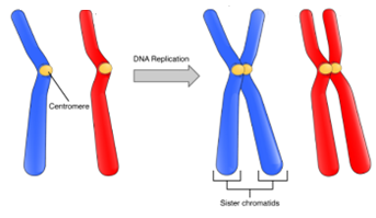
(a)
Figure 8.1: (a) Phases of the Cell Cycle of Somatic cells; (b) In the S phase, DNA is duplicated to prepare for mitosis, where the duplicated DNA forms two sister chromatids held together by a centromere. The sister chromatids will separate during mitosis and be distributed to the two daughter cells.
Note to students: Write all data and answers to questions on the Lab Report provided.
Activity 1: Match the terms of the cell cycle with their descriptions.
|
Term |
Definition |
|
a. Cell Cycle |
1. Body cell that forms tissues, organs, and structures |
|
b. Mitosis |
2. Nuclear division of somatic cells that results in 2 diploid daughter cells |
|
c. Meiosis |
3. Cytoplasmic division |
|
d. Cytokinesis |
4. Reproductive cell or gamete |
|
e. S phase |
5. A series of ordered events that leads to cell division; has two phases: interphase and M phase |
|
f. G1 and G2 phases |
6. Phase of the cell cycle where DNA is replicated |
|
g. Somatic cell |
7. Nuclear division of germ cells that results in 4 haploid daughter cells |
|
h. Germ Cell |
8. Phases of the cell cycle where growth and replication of organelles occur |
MITOSIS
Mitosis is a type of nuclear division where one somatic cell is divided into two genetically identical somatic daughter cells. It is essential for growth, development, and repair of cells. It includes 4 phases: prophase, metaphase, anaphase, and telophase, where chromosomes condense, line up, and separate into two daughter cells. After nuclear division is complete, the cell must go through cytokinesis where the cytoplasm of the parent cell splits into two daughter cells. (Figure 8.2)
-
- Prophase: Genetic material in the form of chromatin fibers condense into chromosomes. Chromosomes are DNA-containing structures consisting of two sister chromatids held together by a centromere. The nuclear envelope starts to break down, and the mitotic spindle begins to form, emerging from the centrioles.
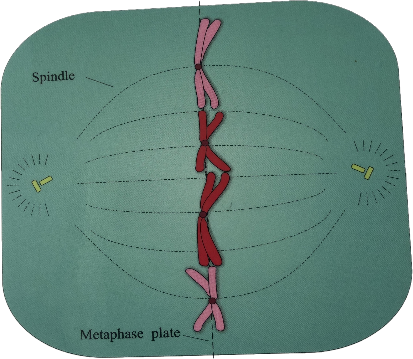
- Metaphase: The nuclear envelope is completely gone. The mitotic spindle fibers bind to the centromeres of the chromosomes to align the chromosomes along the equator of the cell, or metaphase plate. All the chromosomes are lined up in the middle of the spindle.
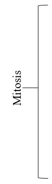
- Anaphase: The sister chromatids are pulled apart by the spindle fibers towards the opposite poles of the cell.
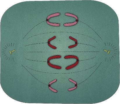
- Telophase: The chromosomes arrive at the two poles and begin to uncoil into chromatin. New nuclear envelopes form around each set of chromosomes, and the spindle fibers break down.
- Cytokinesis: The cytoplasm divides, creating two separate identical daughter cells. In animal cells this occurs through the formation of a cleavage furrow.
 Figure 8.2: M phase of the Cell Cycle consists of nuclear division (mitosis), followed by Cytokinesis.
Activity 2: Identify the Phases of Mitosis
a. Label the image with the proper phase of mitosis. Record your answers on the Lab Report.
b. Number the images of the phases in proper sequence of mitosis. Record your answer on the Lab Report.
1. 2.
2.  3.
3.  4.
4.
MEIOSIS
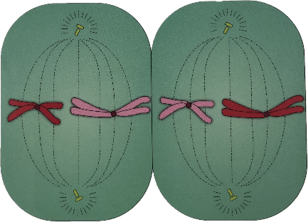
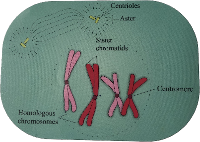
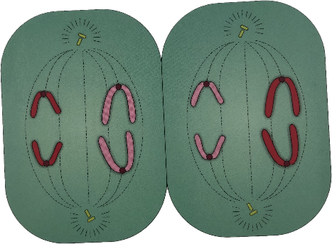
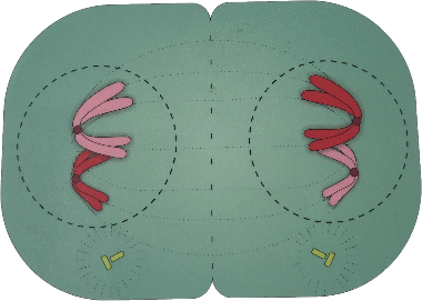
Meiosis is a type of nuclear division of germ cells where the chromosome number is reduced by half producing gametes that are haploid. For example, in humans, the chromosome number is reduced from 46 to 23. This occurs due to meiosis occurring in two successive stages: meiosis I and meiosis II. The phases within meiosis I and meiosis II are named in a similar manner to mitosis: prophase, metaphase, anaphase, and telophase. After each stage of meiosis, cytokinesis takes place (Figure 8.3) Meiosis I involves the separation of homologous chromosome pairs, and meiosis II, the separation of sister chromatids, (In mitosis, only one cell division occurs involving the separation of sister chromatids.) Homologous chromosomes are pairs of chromosomes found in diploid cells, each inherited from one parent. (There are one maternal and one paternal chromosome in each pair). Independent assortment, that is the separation of chromosomes during meiosis, produces new combinations and tremendous variation in gametes. Each homologous chromosome pair acts independently of each other as they align along the metaphase plate and separate randomly, ensuring that each daughter cell receives one chromosome from each pair, with no predictable pattern. (Figure 8.5).
During prophase I, additional genetic variation is introduced due to crossing over between non-sister chromatids of homologous chromosomes (Figure 8.4). Crossing over involves the breaking and reattachment of segments of homologous chromosomes to make new combinations of genes in the offspring.
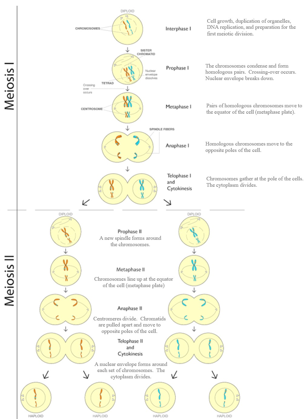
Figure 8.3: Meiosis consists of two stages: meiosis I and meiosis II. Meiosis begins with one diploid parent cell and results in 4 haploid daughter cells.
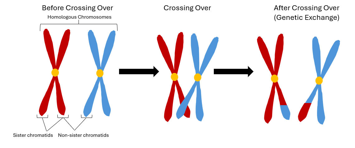
Figure 8.4: Crossing over introduces genetic variation during prophase I.
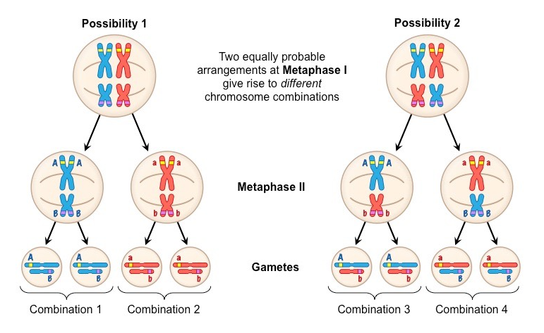 Figure 8.5: The orientation of each homologous pair is random and is not affected by the orientation of any other homologous pair. This gives rise to different chromosome combinations.
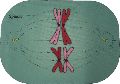
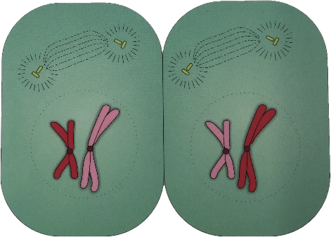
Activity 3: Identify the Phase of Meiosis
a. Identify the phase of meiosis for each image. Record your answers on the Lab Report.
b. Number the images of the phases in proper sequence of meiosis. Record your answer on the Lab Report.
 2.
2.  3.
3.  4.
4. 
5.  6.
6.  7.
7. 8.
8.
GAMETOGENESIS
Gametogenesis is the production of haploid cells through meiosis, resulting in gametes (spermatozoa and oocytes). The production of spermatozoa is called spermatogenesis, and the production of oocytes is called oogenesis. While both processes involve meiosis, they differ in the number of viable haploid cells and the distribution of cytoplasm and organelles. In spermatogenesis, cytoplasmic division is equal, and each diploid cell yields four viable haploid sperm cells. However, in oogenesis, the cytoplasm is unevenly distributed, resulting in one large viable haploid egg cell and three smaller, non-viable polar bodies. (Figure 8.6)
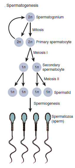
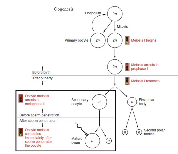
Figure 8.6: Comparison of Spermatogenesis and Oogenesis. Spermatogenesis results in 4 viable haploid sperm cells whereas oogenesis results in 1 viable haploid egg cell and 3 non-viable polar bodies.
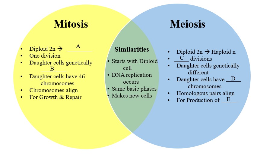
Activity 4: Compare Mitosis and Meiosis
Fill in the missing information in the Venn Diagram that compares the processes of mitosis and meiosis. Record your answers on the Lab Report.
Activity 5: Lab Review
On the Lab Report, answer the questions in the Lab Review section.
Link to Lab Report: Lab 8 Cell Reproduction Lab Report
REFERENCES
Betts, J. G., Young, K. A., Wise, J. A., Johnson, E., Poe, B., Kruse, D. H., Korol, O., Johnson, J. E., Womble, M., & DeSaix, P. (2022a, April 20). 3.5 Cell Growth and Division – Anatomy and Physiology 2E | OpenStax. https://openstax.org/books/anatomy-and-physiology-2e/pages/3-5-cell-growth-and-division
Betts, J. G., Young, K. A., Wise, J. A., Johnson, E., Poe, B., Kruse, D. H., Korol, O., Johnson, J. E., Womble, M., & DeSaix, P. (2022d, April 20). Ch. 27 Introduction – The Reproductive System | OpenStax. https://openstax.org/books/anatomy-and-physiology-2e/pages/27-introduction
BioCam. (n.d.). Biology Wall Charts & Dissection Charts. https://www.biocam.com/
BioNinja. (n.d.). Random Assortment. https://old-ib.bioninja.com.au/higher-level/topic-10-genetics-and-evolu/101-meiosis/random-assortment.html
Bush, Patricia. (2025). Genetic Variation: Crossing Over. Wayne, NJ: William Paterson University.
Koczwara, Katherine, “Meiosis”. Embryo Project Encyclopedia ( 2022-01-21 ). ISSN: 1940-5030 https://keep.lib.asu.edu/items/175307
Mader, Sylvia S. (2023). Laboratory Manual for Human Biology. 17th edition. McGraw-Hill.
Mukherjee, S. (2023, February 2). Cell Cycle: Definition, Phases, and diagram. Science Facts. https://www.sciencefacts.net/cell-cycle.html
Tortora, Gerard J. and Bryan H. Derrickson. (2020). Principles of Anatomy and Physiology, 16th edition. John Wiley and Sons.
